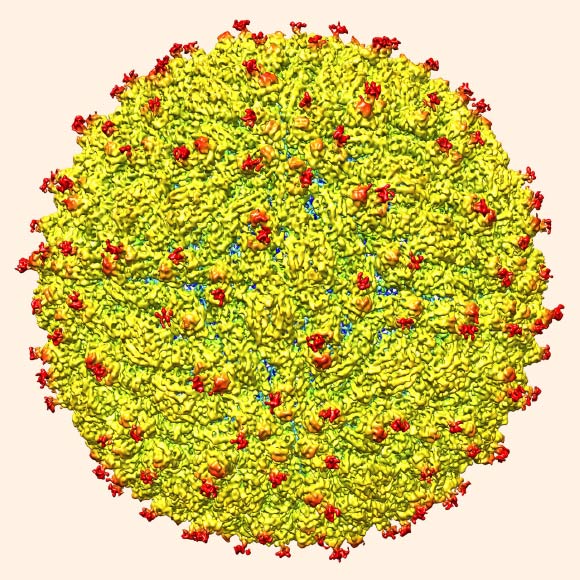
A team of researchers led by Purdue University scientists Michael Rossmann and Richard Kuhn is the first to determine the structure of the Zika virus, which reveals insights critical to the development of effective antiviral treatments and vaccines. A paper detailing the findings was published online this week in the journal Science.

A representation of the surface of Zika virus with protruding envelope glycoproteins (red) shown. Image credit: Devika Sirohi et al / Purdue University.
Zika virus is an emerging mosquito-borne flavivirus closely related to dengue virus. It was first identified in a rhesus monkey from Zika forest in Uganda in 1947 and in mosquitoes Aedes africanus in the same forest in 1948. It was subsequently identified in humans in 1952 in Uganda and Tanzania.
Outbreaks of Zika virus disease have been recorded in Africa, the Americas, Asia and the Pacific.
Zika virus is transmitted to people through the bite of an infected mosquito from the Aedes genus, mainly Aedes aegypti in tropical regions. This is the same mosquito that transmits dengue, chikungunya and yellow fever.
Some evidence suggests the virus can also be transmitted to humans through blood transfusion, perinatal transmission and sexual transmission. However, these modes are very rare. For example, sexual transmission of Zika virus has been described in two cases, and the presence of the Zika virus in semen in one additional case.
The incubation period of Zika virus is typically between 2 and 7 days. Infection is characterized by low grade fever (less than 101.3 degrees Fahrenheit, or 38.5 degrees Celsius) frequently accompanied by a maculopapular rash.
Other common symptoms include muscle pain, joint pain with possible swelling, headache, pain behind the eyes and conjunctivitis. As symptoms are often mild, infection may go unrecognized or be misdiagnosed as dengue.
The ongoing Zika virus epidemic is of special concern because of apparent links to congenital microcephaly, a medical condition in which the brain does not develop properly, resulting in a smaller than normal head, as well as the autoimmune-neurological Guillain Barre syndrome.
To learn more about the structure of the virus and possible ways to therapeutically target it, Dr. Rossmann, Dr. Kuhn and their colleagues used cryo-electron microscopy to analyze a strain isolated from an infected patient during the French Polynesia epidemic in 2013-14.
“We were able to determine through cryo-electron microscopy the virus structure at a resolution that previously would only have been possible through X-ray crystallography,” Dr. Rossmann said.
The analysis reveals that the structure of Zika virus is very similar to that of other flaviviruses, and particularly similar to dengue.
The scientists also identified regions within the virus structure where it differs from other flaviviruses.
According to the team, the difference is in a region of the E glycoprotein that flaviviruses may use to attach to some human cells.
The variation in the E glycoprotein of Zika virus could explain the ability of the virus to attack nerve cells, as well as the associations of Zika virus infection with birth defects and the Guillian-Barre syndrome.
The structure could inform vaccine development, as the Zika E glycoprotein is a key target of immune responses against the virus.
The information may also be useful for designing treatments such as antiviral drugs or antibodies that interfere with E glycoprotein function.
Further, details on the structural differences between the Zika E glycoprotein and the same protein in dengue virus may make it possible to create diagnostic tests that can distinguish Zika virus infection from dengue infection, a critical need in countries where both Zika and dengue viruses are circulating.
_____
Devika Sirohi et al. The 3.8 Å resolution cryo-EM structure of Zika virus. Science, published online March 31, 2016; doi: 10.1126/science.aaf5316


