An international team of scientists led by Museums Victoria and the University of Melbourne has scanned all known joey specimens of the Tasmanian tiger (Thylacinus cynocephalus) — an iconic Australian marsupial predator that was hunted to extinction in the early 1900s — to create 3D digital models which have allowed them to reconstruct their growth and development. The digital scans show that when first born the Tasmanian tiger looked like any other marsupial; but three months later, when they left the pouch they had taken on the appearance of a puppy and continued to grow with a dog-like appearance. The findings are published in the journal Royal Society Open Science.

A Tasmanian tiger (Thylacinus cynocephalus) with three cubs at Beaumaris Zoo in Hobart, 1909. Image credit: Tasmanian Musuem and Art Gallery.
The Tasmanian tiger — otherwise known as the thylacine — was a large Australian marsupial mammal known from the island state of Tasmania.
Once ranging throughout Australia and New Guinea, the species disappeared from the mainland around 3,000 years ago, probably through competition and predation by humans and dingoes. A remnant population became isolated on Tasmania before they were hunted to extinction in the early twentieth century, with the last known individual dying in captivity in Hobart Zoo in 1936.
Its resemblance to the dingo is one of the best examples of ‘convergent evolution’ in mammals. This is where, two species, despite not being closely related, evolve to look very similar.
The Tasmanian tiger would have last shared a common ancestor with the canids (dogs and wolves) around 160 million years ago.
“After sequencing the Tasmanian tiger genome in 2017, this new research fills one more piece of the puzzle on why they have evolved to look so similar to dogs,” said Dr. Christy Hipsley, from Museums Victoria and the University of Melbourne.
“This is the first digital development series of the Tasmanian tiger. Using CT technology we have been able to garner new information on the biology of this iconic species, and its growth and development.”
“These scans show in incredible detail how the Tasmanian tiger started its journey in life as a joey that looked very much like any other marsupial, with robust forearms so that it could climb into its mothers pouch,” Dr. Hipsley said.
“But by the time it left the pouch around 12 weeks to start independent life, it looked more like a dog or wolf, with longer hind limbs than forelimbs.”
“Until now, there have only been limited details on its growth and development. For the very first time we have been able to look inside these remarkably rare and precious specimens,” said Ph.D. student Axel Newton, also from Museums Victoria and the University of Melbourne.

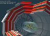
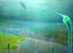
Soft tissue and skeletal reconstructions of the Tasmanian tiger: (a) lateral view of the 9.5-week-old Tasmanian tiger with semi-transparent skin, internal organs and bony elements. The brain (orange), trachea (green), lungs (yellow), heart (red), liver (purple) and kidneys (pink) were separately rendered and overlaid. Teeth showed the highest density (white) followed by the bones of the cranium and limb bones. Lighter elements, including the skin, appear translucent; (b) lateral view of the reconstructed pouch young. Abbreviations: as - axial spines, at - atlas, ax - axis, bc - bronchioles, br - brain, ca - calcaneus, cl - clavicles, ht - heart, il - ilium, is - ischium, ki - kidney, lu - lung, lvr - liver, ms - maxillary swellings, sc - scapula, st - sternum, tr - trachea. Image credit: Newton et al, doi: 10.1098/rsos.171914.
Unable to study the living species, the researchers had to look to the 13 Tasmanian tiger joey specimens that exist in museum collections worldwide.
These joey specimens, representing five stages of postnatal development, were scanned using non-invasive X-ray micro-CT scanning technology to create high resolution 3D digital models, in which all their internal structures such as skeleton and organs could be studied.
“This was an incredibly effective technique to study the skeletal anatomy of the specimens without causing any damage to them,” said Dr. Andrew Pask, also from the University of Melbourne and Museums Victoria.
“This research clearly demonstrates the power of CT technology. It has allowed us to scan all the known Tasmanian tiger joey specimens in the world, and study their internal structures in high resolution without having to dissect or cause damage to the specimen.”
“By examining their bone development, we’ve been able to illustrate how the Tasmanian tiger matured and identify when they took on the appearance of a dog.”
The study also revealed the incorrect classification of two specimens held in the collection of the Tasmanian Museums and Art Gallery. Instead, they are most likely to be quolls or Tasmanian devils, based on the number of vertebrate and presence of large epipubic bones (specialized bones that support the pouch in modern marsupials).
The 3D digital Tasmanian tiger models are publicly available as a resource for current and future researchers.
_____
Axel H. Newton et al. 2018. Letting the ‘cat’ out of the bag: pouch young development of the extinct Tasmanian tiger revealed by X-ray computed tomography. R. Soc. open sci 5 (2): 171914; doi: 10.1098/rsos.171914