The Brain Atlas explores the protein expression in the mammalian brain by visualization and integration of data from three species of mammals: human, pig and mouse. The open-access resource is based on the analysis of nearly 1,900 brain samples covering 27 brain regions.

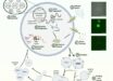
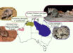
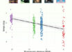
Genome-wide transcriptomics analysis of anatomically dissected regions in mammalian brains uncovers regional and species-specific expression: multiple regions of the human, pig, and mouse brain were dissected and analyzed; a uniform manifold approximation and projection (UMAP) analysis (middle) shows the global expression patterns of 1,710 samples in the human brain, with the cerebellum as the outlier; the Human Protein Atlas (HPA) Brain Atlas (right) shows the expression of individual genes, for example, synaptosomal-associated protein 25 (SNAP25), in the different brain regions in the three mammalian species. Image credit: Sjöstedt et al, doi: 10.1126/science.aay5947.
“As expected the blueprint for the brain is shared among mammals, but the new map also reveals interesting differences between human, pig and mouse brains,” said Professor Mathias Uhlén, a researcher in the Department of Protein Science at KTH Royal Institute of Technology and the Department of Neuroscience at Karolinska Institutet and Director of the Human Protein Atlas effort.
The cerebellum emerged in the study as the most distinct region of the brain.
Many proteins with elevated expression levels in this region were found, including several associated to psychiatric disorders supporting a role of the cerebellum in the processing of emotions.
“Another interesting finding is that the different cell types of the brain share specialized proteins with peripheral organs,” said Dr. Evelina Sjöstedt, a scientist in the Department of Neuroscience at Karolinska Institutet.
“For example, astrocytes, the cells that ‘filter’ the extracellular environment in the brain share a lot of transporters and metabolic enzymes with cells in the liver that filter the blood.”
When comparing the neurotransmitter systems, responsible for the communication between neurons, some clear differences between the species could be identified.
“Several molecular components of neurotransmitter systems, especially receptors that respond to released neurotransmitters and neuropeptides, show a different pattern in humans and mice,” said Dr. Jan Mulder, a researcher in the Department of Neuroscience at Karolinska Institutet.
“This means that caution should be taken when selecting animals as models for human mental and neurological disorders.”
For selected genes/proteins, the Brain Atlas also contains microscopic images showing the protein distribution in human brain samples and detailed, zoomable maps of protein distribution in the mouse brain.
The results were published in the March 6, 2020 issue of the journal Science.
_____
Evelina Sjöstedt et al. 2020. An atlas of the protein-coding genes in the human, pig, and mouse brain. Science 367 (6482): eaay5947; doi: 10.1126/science.aay5947