According to a group of researchers led by Prof Susumu Tonegawa from Massachusetts Institute of Technology, memories that have been lost as a result of amnesia can be recalled by activating brain cells with a technology known as optogenetics.
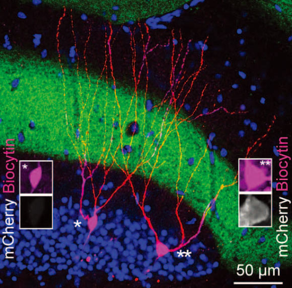
Image of a pair of engram (mCherry+) and nonengram (mCherry–) cells in the hippocampal dentate gyrus. Image credit: Tomás J. Ryan et al.
Scientists have for many years debated whether retrograde amnesia – a condition associated with traumatic brain injury, Alzheimer’s disease, and other neurological disorders – is caused by damage to specific brain cells, meaning a memory cannot be stored, or if access to that memory is somehow blocked, preventing its recall.
“The majority of scientists have favored the storage theory, but we have shown in the study that this majority theory is probably wrong. Amnesia is a problem of retrieval impairment,” Prof Tonegawa said.
Researchers have previously speculated that somewhere in the brain network is a population of neurons that are activated during the process of acquiring a memory, causing enduring physical or chemical changes.
If these groups of neurons – known as memory engram cells – are subsequently reactivated by a trigger such as a particular sight or smell, for example, the entire memory is recalled.
In a 2012 study, Prof Tonegawa and co-authors used optogenetics – in which proteins are added to neurons to allow them to be activated with light – to demonstrate that such a population of neurons does indeed exist in an area of the brain called the hippocampus. However, until now no one has been able to show that these groups of neurons do undergo enduring chemical changes, in a process known as memory consolidation.
In the new study, the researchers demonstrated in mice that traces of old memories do remain in the amnesic brain, and that the cellular pathways underlying them can be reactivated, allowing ‘lost’ memories to be found.
To make mice amnesic, they trained the animals to associate a mild foot shock with a specific environment, chamber A, eliciting a typical ‘freezing’ behavior.
“Eventually, trained mice would freeze in chamber A even without the shock. Neurons activated during memory formation were genetically labeled to allow their visualization and reactivation.”
Then, some mice were given anisomycin, an antibiotic produced by Streptomyces griseolus which inhibits protein synthesis and prevents increases in synaptic strength important for memory encoding, thus inducing retrograde amnesia. Other mice received saline as a control.
“As expected, amnesic mice returned to chamber A did not freeze, indicating that they could not recall the memory for the specific association of the chamber and the mild foot shock.”
Next, to investigate whether the stored memory from the foot shock training in chamber A was absent from the amnesic mice or remained present but was not retrievable, the scientists used optogenetics to selectively activate neurons that were genetically labeled during their training in chamber A with a blue light-sensitive protein, channelrhodopsin, but this time while the mice were in a novel, neutral environment, chamber B.
During activation of the cells involved in the foot shock memory with blue light pulses, the amnesic mice froze just as much as the control mice, indicating that they remembered that they had acquired the memory, even though they could not recall it when placed in chamber A.
To explain how the ‘lost’ memory was recalled during light stimulation of the memory engram, despite the induction of retrograde amnesia, the scientists suggest that different processes may control memory encoding and recall.
For example, during the training period, brain connections between unique memory engrams in neighboring brain structures may be strengthened and once this has occurred, may not require an increase in synaptic strength in order to store, but not recall, the contextual fear memory and would be preserved in the amnesic state.
Indeed, they observed that connectivity was enhanced between memory engram cells in the fear memory-holding amygdala and context memory-holding hippocampus of amnesic mice, even though synaptic changes remained stable.
“Our conclusion is that in retrograde amnesia, past memories may not be erased, but could simply be lost and inaccessible for recall,” said Prof Tonegawa, who is the senior author of the paper reporting the results in the journal Science.
“These findings provide striking insight into the fleeting nature of memories, and will stimulate future research on the biology of memory and its clinical restoration.”
_____
Tomás J. Ryan et al. 2015. Engram cells retain memory under retrograde amnesia. Science, vol. 348, no. 6238, pp. 1007-1013; doi: 10.1126/science.aaa5542