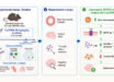
Does a given color elicit comparable neural activity in two different observers? Do colors elicit area-specific response patterns? To address these questions, a duo of neuroscientists at the University of Tübingen predicted what color someone is seeing based on their brain activity, using only knowledge of color responses from other observers’ brains. Estimating the commonalities across brains in the way they respond to achromatic, spatial stimulation allowed the authors to retinotopically align different brain responses to each other in a common response space. In this space derived without any color responses, they could decode across individuals what color an observer was seeing and found spatial color biases that differed between areas.

Using a sample of male and female volunteers, Michael M. Bannert & Andreas Bartels asked whether spatial color biases are shared across different human observers, and whether they are idiosyncratic for distinct areas. Image credit: Vat Loai.
Using functional MRI scanning, University of Tübingen researchers Michael Bannert and Andreas Bartels recorded images from the brains of subjects who were observing visual stimuli, and identified different signals for red, green and yellow.
The pattern of brain activity appeared similar in subjects who were not previously involved, meaning that the color they saw could be correctly predicted simply by comparison with images from the brains of other participants.
The representation of colors in the brain is far more uniform across individuals than had been thought.
It was already possible to determine what color an individual is seeing in experiments using functional magnetic resonance imaging (fMRI), but only for the same brain.
“We wanted to know though how similar colors are encoded in different brains,” Dr. Bannert said.
“In other words, can the colors that are seen also be deduced if we only have the neuronal color signals from the brains of other people?”
“It is well known that the functional structure of different brains is roughly the same.”
“Some regions, for example, are more active when we see a face, a body or just color.”
In their experiments with color rings, the researchers trained specific classification algorithms with fMRI data to distinguish the signals from the brains of a group of individuals systematically by color.
In the next step, they worked with data from new subjects to determine which colors they were seeing using neuronal signals.
To provide orientation in each brain, the scientists spatially mapped how it responded to stimulation at different locations of the visual field using fMRI measurements.
“In order not to bias our results, we didn’t use any colors at this stage, only black & white patterns,” Professor Bartels said.
“Just by using this mapping data in combination with the color information from the brains of others, we were able to determine reliably from the activity of a ‘new’ brain which colors that person was seeing at that point.”
“We were surprised that even the subtle differences between individual colors are so similar across brains in terms of the activity patterns they elicit during processing in certain visual areas. This was not previously known.”
The spatial color coding in the brain is area-specific and organized consistently across individuals.
“There must be a functional or evolutionary pressure for this uniform development, but this relationship still needs to be clarified,” the authors said.
The study was published this week in the Journal of Neuroscience.
_____
Michael M. Bannert & Andreas Bartels. Large-scale color biases in the retinotopic functional architecture are region specific and shared across human brains. Journal of Neuroscience, published online September 8, 2025; doi: 10.1523/JNEUROSCI.2717-20.2025