An international team of paleoanthropologists led by the University of the Witwatersrand has examined the first cervical vertebra (atlas) of the ‘Little Foot,’ a 3.67 million-year-old Australopithecus prometheus specimen from the Sterkfontein cave in South Africa comprised of a skull and associated postcranial skeleton.

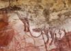
The Little Foot fossil in the Sterkfontein cave, central South Africa. Image credit: Purdue University.
“The atlas plays a crucial role in vertebrate biology,” said Dr. Amélie Beaudet from the School of Geography, Archaeology and Environmental Studies at the University of the Witwatersrand and the Department of Anatomy at the University of Pretoria and colleagues.
“Besides acting as the connection between the head and the neck, the atlas also plays a role in how blood is supplied to the brain via the vertebral arteries.”
The researchers compared the atlas of the Little Foot (also known as StW 573) with other Australopithecus fossils from Africa as well as humans, chimpanzees, gorillas and orangutans.
They found that Australopithecus prometheus was capable of head movements that differ from modern humans.
“The morphology of the first cervical vertebra, or atlas, reflects multiple aspects of an organism’s life,” Dr. Beaudet said.
“In particular, the nearly complete atlas of the Little Foot has the potential to provide new insights into the evolution of head mobility and the arterial supply to the brain in the human lineage.”
The shape of the atlas determines the range of head motions while the size of the arteries passing through the vertebrae to the skull is useful for estimating blood flow supplying the brain.
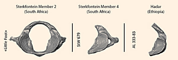
Comparison of the nearly intact first cervical vertebra of the Little Foot and two Australopithecus from Sterkfontein in South Africa and from Hadar in Ethiopia. Image credit: Amélie Beaudet / Wits University.
“Our study shows that Australopithecus prometheus was capable of head movements that differ from us,” Dr. Beaudet said.
“This could be explained by the greater ability of Australopithecus prometheus to climb and move in the trees.”
“However, a southern African Australopithecus specimen younger than the Little Foot (probably younger by about 1 million years) may have partially lost this capacity and spent more time on the ground, like us today.”
The overall dimensions and shape of the atlas of the Little Foot are similar to living chimpanzees.
More specifically, the ligament insertions — that could be inferred from the presence and configuration of bony tubercles — and the morphology of the facet joints linking the head and the neck all suggest that Australopithecus prometheus was moving regularly in trees.
The team also demonstrated that blood flow in the Little Foot, and thus the utilization of glucose by the brain, was about three times lower than in living humans, and closer to those of living chimpanzees.
“The low investment of energy into the brain of Australopithecus prometheus could be tentatively explained by a relatively small brain of the specimen (around 408 cm3), a low quality diet (low proportion of animal products) or high costs of other aspects of the biology of Australopithecus prometheus (such as upright walking),” Dr. Beaudet said.
“In any case, this might suggest that the human brain’s vascular system emerged much later in our history.”
The results appear in the journal Scientific Reports.
_____
A. Beaudet et al. 2020. The atlas of StW 573 and the late emergence of human-like head mobility and brain metabolism. Sci Rep 10, 4285; doi: 10.1038/s41598-020-60837-2