A team of paleontologists co-led by Drs Sergio Bertazzo and Susannah Maidment of Imperial College London has discovered what look like remnants of red blood cells and collagen fibers in 75-million-year-old dinosaur specimens.
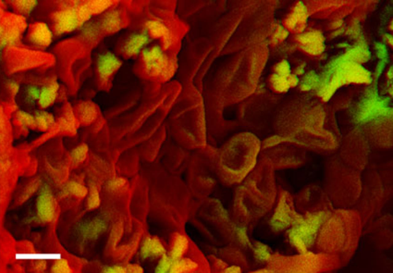
Density-dependent color scanning electron micrograph of an ungual claw of an indeterminate theropod dinosaur shows erythrocyte-like structures. Scale bar – 1 μm. Image credit: Sergio Bertazzo et al.
In their study, the paleontologists analyzed eight fragments of dinosaur bones (Chasmosaurus sp.; ceratopsid, hadrosaurid and theropod dinosaurs) that have for more than a century been in the collections of Natural History Museum in London.
They used a scanning electron microscopy device to observe the structure, composition and location of the soft tissue inside the dinosaur fossil fragments. They then used a focused ion beam to slice into the samples and observe the internal structure of the fossils.
The scientists examined part of a theropod dinosaur claw and identified tiny structures that look ovoid and with an inner denser core.
These could potentially be red blood cells although the paleontologists caution that further evidence would be needed to confirm that the structures do not have another origin. The hope is that if red blood cells can be found in fossilized dinosaur fragments, this could help scientists to understand when dinosaurs evolved a warm blooded, bird-like metabolism.
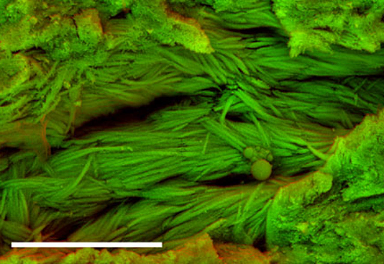
Density-dependent color scanning electron micrograph of a rib fragment of an indeterminate dinosaur shows fibrous structures. Scale bar – 5 μm. Image credit: Sergio Bertazzo et al.
Dr Bertazzo, Dr Maidment and their colleagues also examined the fossils using a transmission electron microscope to detect the fibrous structures.
In one dinosaur fossil fragment, they found structures that looked fibrous and had a banded structure similar to the banding that can be seen in modern day collagen fibers.
Further evidence would be needed to definitively conclude that the structures found originate from a preservation of collagen. If verified, the identification of collagen-like structures could in the future provide a new independent line of evidence to show how various dinosaur groups are related to each other.
“We still need to do more research to confirm what it is that we are imaging in these dinosaur bone fragments, but the ancient tissue structures we have analyzed have some similarities to red blood cells and collagen fibers. If we can confirm that our initial observations are correct, then this could yield fresh insights into how these creatures once lived and evolved,” said Dr Bertazzo, lead author on the study published in the journal Nature Communications.
“Our study is helping us to see that preserved soft tissue may be more widespread in dinosaur fossils than we originally thought,” Dr Maidment added.
“Although remnants of soft tissues have previously been discovered in rare, exceptionally preserved fossils, what is particularly exciting about our study is that we have discovered structures reminiscent of blood cells and collagen fibers in scrappy, poorly preserved fossils. This suggests that this sort of soft tissue preservation might be widespread in fossils.”
_____
Sergio Bertazzo et al. 2015. Fibres and cellular structures preserved in 75-million–year-old dinosaur specimens. Nature Communications 6, article number: 7352; doi: 10.1038/ncomms8352