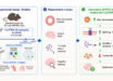
A team of neuroscientists at Howard Hughes Medical Institute’s Janelia Research Campus has taken detailed pictures of the whole brain of an adult female fruit fly (Drosophila melanogaster) using electron microscopy.
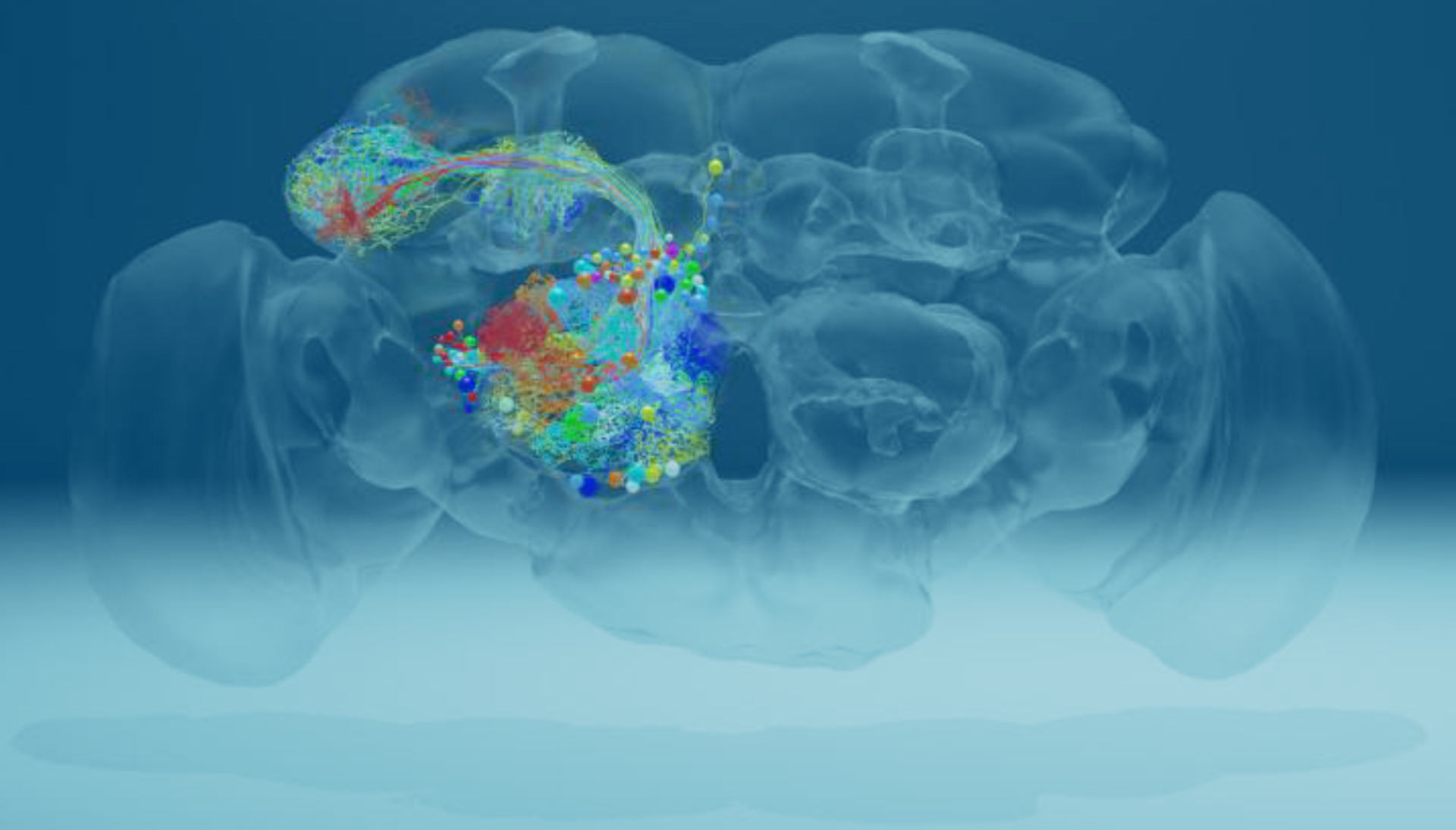
The fruit fly brain contains roughly 100,000 neurons, which can now be traced in detail using a dataset that includes roughly 21 million images Zheng et al traced the paths of neurons (colored threads) that reach out to the mushroom body, a region involved in memory and learning. Image credit: Zheng et al, doi: 10.1016/j.cell.2018.06.019.
The fruit fly brain contains about 100,000 neurons. Each neuron branches into a starburst of fine wires that touch the wires of other neurons. Neurons talk to one another through these touchpoints, or synapses, forming a dense mesh of communication circuits.
Scientists can view these wires and synapses with an imaging technique called serial section transmission electron microscopy.
First, they infuse the fly’s brain with a cocktail of heavy metals. These metals pack into cell membranes and synapses, ultimately marking the outlines of each neuron and its connections.
Then they hit slices of the brain with a beam of electrons, which passes through everything except the metal-loaded parts.
“The entire fly brain has never been imaged before at this resolution that lets you see connections between neurons,” said Dr. David Bock, senior author of the study.
“That detail is key for mapping out the brain’s circuitry — the precise webs of neuronal connections that underpin specific fly behaviors.”
“Now we can trace the path of any one neuron to any other neuron throughout the whole brain.”
The millions of images Dr. Bock and colleagues collected and stitched together offer an in-depth look at the fly brain — and the chance to explore uncharted areas.
The researchers traced the paths of neurons that reach out to the mushroom body, a region involved in memory and learning.
These cells, called olfactory projection neurons, have been well described previously, using light microscopy.
Manually tracing the outlines of these neurons and all their wirelike projections let the team confirm the quality of their image data.
“We stubbed our toe on some interesting new things as well,” Dr. Bock said.
Olfactory projection neurons send messages to neurons called Kenyon cells. These cells, in turn, talk to different sets of neurons. Until now, scientists hadn’t identified Kenyon cells’ conversation partners in a region of the mushroom body called the calyx.
Dr. Bock and co-authors pinpointed some of these neurons, as well as a previously unknown brain-spanning neuron that also relays information to Kenyon cells.
“The olfactory projection neurons also appeared to be more tightly bundled together than researchers had thought,” Dr. Bock said.
“This bundling suggests an orderly structure in something once believed to be largely random.”
“A better understanding of this brain circuitry could give scientists insight into fly behavior,” he added.
“We think it will tell us something about how the animal learns — how it associates odors with a reward or punishment.”
The results were published in the journal Cell.
_____
Zhihao Zheng et al. A Complete Electron Microscopy Volume of the Brain of Adult Drosophila melanogaster. Cell, published online July 19, 2018; doi: 10.1016/j.cell.2018.06.019