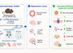
A team of neuroscientists at the University of Southern California Mark and Mary Stevens Neuroimaging and Informatics Institute has produced the most detailed atlas yet of the hippocampus — the brain’s memory bank.
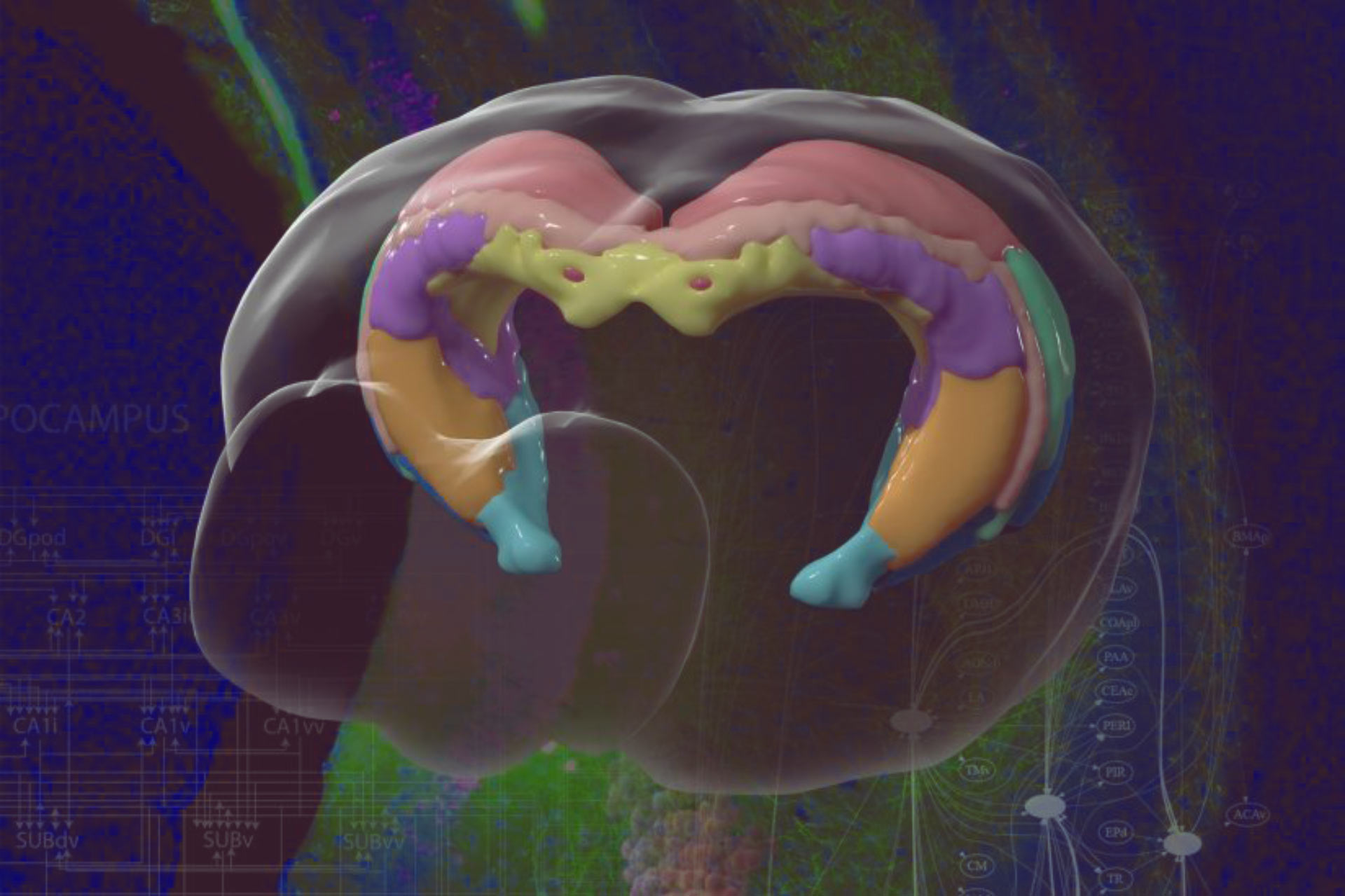
Brain mapping helps neuroscientists understand how specific areas function and how to create new drugs and targeted treatments. Image credit: Jim Stanis and Tyler Ard, USC Mark and Mary Stevens Neuroimaging and Informatics Institute.
The human hippocampus sits at the base of the brain. It stores memories, helps regulate emotions and guides navigation by spatial processing.
It’s the first part of the brain impaired by Alzheimer’s; hippocampus degeneration also can cause epilepsy and other diseases.
“Like a new atlas, we’ve constructed the most detailed diagram of the hippocampus to date. With a better map, we can see each region and how it functions,” said Dr. Michael Bienkowski, co-lead author of the study.
“A better map is a resource scientists can use to better understand the hippocampus and how its degeneration leads to diseases.”
“Researchers can use the new map of the hippocampus to deliver genetically targeted drugs to specific neurons with fewer side effect,” said Dr. Hong-Wei Dong, co-lead author of the study.
Dr. Bienkowski, Dr. Dong and co-authors worked on a mouse brain because it’s organized similar to a human brain.
The work is part of the Mouse Connectome Project, an effort that collects data about neural connections in the brain, sharing it publicly with researchers in more than 100 countries.
“Scientists have known the basic four-part architecture of the hippocampus for a long time,” they said.
“What’s different now is we can show its sub-regions and how nerve cells interact across the structure.”
“It’s a night-and-day difference, akin to seeing transmission lines and power poles slung across a city by day compared to fully illuminated at night.”
“This new visualization traces neural pathways and connections in remarkable detail using fluorescent dyes as tracers that reveal cells, neuron junctions and connections to the rest of the brain.”
“It totally changes our understanding by combining a wiring diagram with gene expression of the mouse hippocampus,” Dr. Bienkowski noted.
“We see it doing different things, and this gives us a new way to understand how the whole thing works together. This should have a very profound and broad impact.”
The team’s results appear in the journal Nature Neuroscience.
_____
Michael S. Bienkowski et al. Integration of gene expression and brain-wide connectivity reveals the multiscale organization of mouse hippocampal networks. Nature Neuroscience, published online October 8, 2018; doi: 10.1038/s41593-018-0241-y