A new study in mice led by Prof Bo Li of the Cold Spring Harbor Laboratory examines how fear responses are learned, controlled, and memorized. The findings show that a particular class of neurons in a subdivision of the amygdala plays an active role in these processes.
")
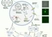
This image shows neurons in the lateral subdivision of the central amygdala, somatostain-positive neurons are shown in red (Bo Li)
Previous studies had indicated that structures inside the amygdalae – a pair of almond-shaped formations that sit deep within the brain and are known to be involved in emotion and reward-based behavior – may be part of the circuit that controls fear learning and memory. In particular, a region called the central amygdala was thought to be a passive relay for the signals relayed within this circuit.
“Neuroscientists believed that changes in the strength of the connections onto neurons in the central amygdala must occur for fear memory to be encoded, but nobody had been able to actually show this,” said Prof Li, who with colleagues reported the findings in the journal Nature Neuroscience.
To examine the behavior of mice undergoing a fear test, the team first trained them to respond in a Pavlovian manner to an auditory cue. The mice began to ‘freeze,’ a very common fear response, whenever they heard one of the sounds they had been trained to fear.
To study the particular neurons involved, and to understand them in relation to the fear-inducing auditory cue, the team used a variety of methods. One of these involved delivering a gene that encodes for a light-sensitive protein into the particular neurons Li’s group wanted to look at.
By implanting a very thin fiber-optic cable directly into the area containing the photosensitive neurons, the scientists were able to shine colored laser light with pinpoint accuracy onto the cells, and in this manner activate them. This is a technique known as optogenetics. Any changes in the behavior of the mice in response to the laser were then monitored.
The ability to probe genetically defined groups of neurons was vital because there are two sets of neurons important in fear-learning and memory processes. The difference between them, the team learned, was in their release of message-carrying neurotransmitters into the spaces called synapses between neurons. In one subset of neurons, neurotransmitter release was enhanced; in another it was diminished. If measurements had been taken across the total cell population in the central amygdala, neurotransmitter levels from these two distinct sets of neurons would have been averaged out, and thus would not have been detected.
The team found that fear conditioning induced experience-dependent changes in the release of neurotransmitters in excitatory synapses that connect with inhibitory neurons – neurons that suppress the activity of other neurons – in the central amygdala. These changes in the strength of neuronal connections are known as synaptic plasticity.
Particularly important in this process were somatostatin-positive (SOM+) neurons. Somatostatin is a hormone that affects neurotransmitter release. Prof Li’s team found that fear-memory formation was impaired when they prevent the activation of SOM+ neurons.
SOM+ neurons are necessary for recall of fear memories, the team also found. Indeed, the activity of these neurons alone proved sufficient to drive fear responses. Thus, instead of being a passive relay for the signals driving fear learning and responses in mice, the team’s work demonstrates that the central amygdala is an active component, and is driven by input from the lateral amygdala, to which it is connected.
“We find that the fear memory in the central amygdala can modify the circuit in a way that translates into action – or what we call the fear response,” Prof Li said.
______
Bibliographic information: Haohong Li et al. Experience-dependent modification of a central amygdala fear circuit. Nature Neuroscience, published online January 27, 2013; doi: 10.1038/nn.3322