A team of researchers from the National Institutes of Health has discovered lymphatic vessels in the dura, the leathery outer coating of the brain. Described in the journal eLife, the discovery holds promise for better understanding the normal physiology of lymphatic drainage from the central nervous system and potential aberrations in neurological diseases.
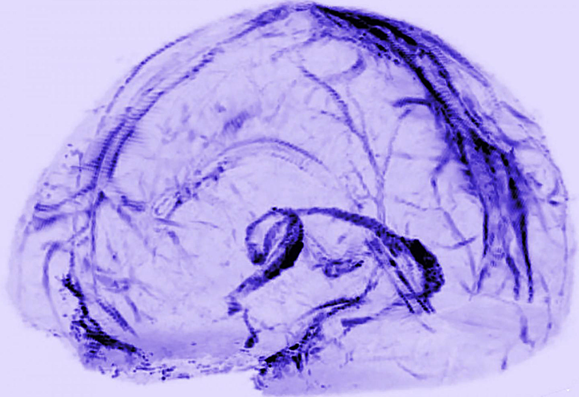
Brain scans of healthy volunteers showed that human brains may drain waste through lymphatic vessels. Image credit: Reich Lab, NIH / NINDS.
Lymphatic vessels are part of the body’s circulatory system. In most of the body they run alongside blood vessels. They transport lymph, a colorless fluid containing immune cells and waste, to the lymph nodes.
Blood vessels deliver white blood cells to an organ and the lymphatic system removes the cells and recirculates them through the body. The process helps the immune system detect whether an organ is under attack from bacteria or viruses or has been injured.
In 1816, an Italian anatomist reported finding lymphatic vessels on the surface of the brain, but for two centuries, it was forgotten.
Until very recently, scientists found no evidence of a lymphatic system in the brain, leaving some puzzled about how the brain drains waste, and others to conclude that brain is an exceptional organ.
Then in 2015, two studies of mice found evidence of the brain’s lymphatic system in the dura.
“I was completely surprised. In medical school, we were taught that the brain has no lymphatic system,” said senior author Dr. Daniel S. Reich, from the NIH’s National Institute of Neurological Disorders and Stroke (NINDS). After those studies, “I thought, maybe we could find it in human brains?”
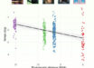
To look for the vessels, Dr. Reich and co-authors used MRI to scan the brains of five healthy volunteers who had been injected with gadobutrol, a magnetic dye typically used to visualize brain blood vessels damaged by diseases, such as multiple sclerosis or cancer.
The dye molecules are small enough to leak out of blood vessels in the dura but too big to pass through the blood-brain barrier and enter other parts of the brain.
At first, when the researchers set the MRI to see blood vessels, the dura lit up brightly, and they could not see any signs of the lymphatic system.
But, when they tuned the scanner differently, the blood vessels disappeared, and the scientists saw that dura also contained smaller but almost equally bright spots and lines which they suspected were lymph vessels.
The results suggested that the dye leaked out of the blood vessels, flowed through the dura and into neighboring lymphatic vessels.
To test this idea, the authors performed another round of scans on two subjects after first injecting them with a second dye made up of larger molecules that leak much less out of blood vessels.
In contrast with the first round of scans, they saw blood vessels in the dura but no lymph vessels regardless of how they tuned the scanner, confirming their suspicions.
They also found evidence for blood and lymph vessels in the dura of autopsied human brain tissue.
Moreover, their brain scans and autopsy studies of brains from nonhuman primates confirmed the results seen in humans, suggesting the lymphatic system is a common feature of mammalian brains.
“These results could fundamentally change the way we think about how the brain and immune system inter-relate,” said Dr. Walter J. Koroshetz, NINDS director.
_____
Martina Absinta et al. 2017. Human and nonhuman primate meninges harbor lymphatic vessels that can be visualized noninvasively by MRI. eLife 6: e29738; doi: 10.7554/eLife.29738