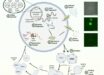
Biologists led by Dr Don Arnold and Dr Richard Roberts from the University of Southern California, Los Angeles, have released an astonishing picture of memories forming inside the mouse brain.

This image shows a living neuron in culture: green dots indicate excitatory synapses; red dots indicate inhibitory synapses (Don Arnold)
The researchers engineered microscopic probes that light up synapses in a living neuron in real time by attaching fluorescent markers onto synaptic proteins. The fluorescent markers allow them to see live excitatory and inhibitory synapses for the first time and, importantly, how they change as new memories are formed. The findings are published in the journal Neuron.
The synapses appear as bright spots along dendrites. As the brain processes new information, those bright spots change, visually indicating how synaptic structures in the brain have been altered by the new data.
“When you make a memory or learn something, there’s a physical change in the brain. It turns out that the thing that gets changed is the distribution of synaptic connections,” Dr Arnold explained.
The probes behave like antibodies, but they bind more tightly and are optimized to work inside the cell – something that ordinary antibodies can’t do.
“Using mRNA display, we can search through more than a trillion different potential proteins simultaneously to find the one protein that binds the target the best,” Dr Roberts said
The probes, named FingRs, are attached to green fluorescent protein, a protein isolated from jellyfish that fluoresces bright green when exposed to blue light. Because FingRs are proteins, the genes encoding them can be put into brain cells in living animals, causing the cells themselves to manufacture the probes.
The design of FingRs also includes a regulation system that cuts off the amount of FingR-GFP that is generated after 100 percent of the target protein is labeled, effectively eliminating background fluorescence – generating a sharper, clearer picture.
These probes can be put in the brains of living mice and then imaged through cranial windows using two-photon microscopy.
______
Bibliographic information: Garrett G. Gross et al. 2013. Recombinant Probes for Visualizing Endogenous Synaptic Proteins in Living Neurons. Neuron, vol. 78, no. 6, pp. 971-985; doi: 10.1016/j.neuron.2013.04.017