In a retrospective case series, an international team of radiologists has reviewed chest CT scans of 21 symptomatic patients from China infected with the 2019-nCoV coronavirus. Typical CT findings included bilateral pulmonary parenchymal ground-glass and consolidative pulmonary opacities, sometimes with a rounded morphology and a peripheral lung distribution. Notably, lung cavitation, discrete pulmonary nodules, pleural effusions, and lymphadenopathy were absent. Follow-up imaging in a subset of patients during the study time window often demonstrated mild or moderate progression of disease as manifested by increasing extent and density of lung opacities.
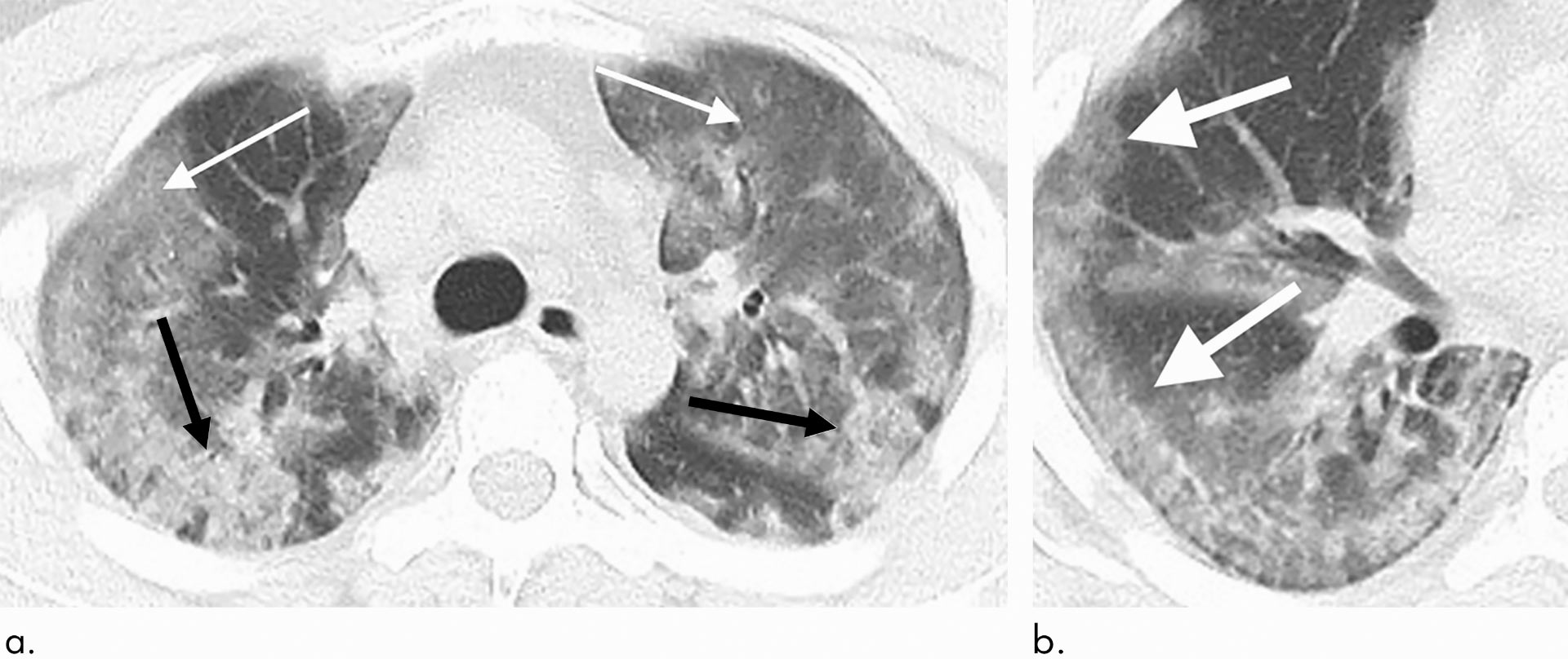
29-year old male with unknown exposure history, presenting with fever and cough, ultimately requiring intensive care unit admission: (a) axial thin-section non-contrast CT scan shows diffuse bilateral confluent and patchy ground-glass (solid arrows) and consolidative (dashed arrows) pulmonary opacities; (b) the disease in the right middle and lower lobes has a striking peripheral distribution (arrow). Image credit: Chung et al, doi: 10.1148/radiol.2020200230.
“Early disease recognition is important not only for prompt implementation of treatment, but also for patient isolation and effective public health surveillance, containment and response,” said Dr. Michael Chung, from the Mount Sinai Health System.
In the study, Dr. Chung and his colleagues set out to characterize the key chest CT imaging findings in a group of patients infected with 2019-nCoV in China with the goal of familiarizing radiologists and clinical teams with the imaging manifestations of this new outbreak.
From January 18, 2020, until January 27, 2020, 21 patients admitted to three hospitals in three provinces in China with confirmed 2019-nCoV infection underwent chest CT.
The 21 patients consisted of 13 men and 8 women ranging in age from 29 to 77 years old, with a mean age of 51.2 years.
All patients were confirmed positive for infection via laboratory testing of respiratory secretions.

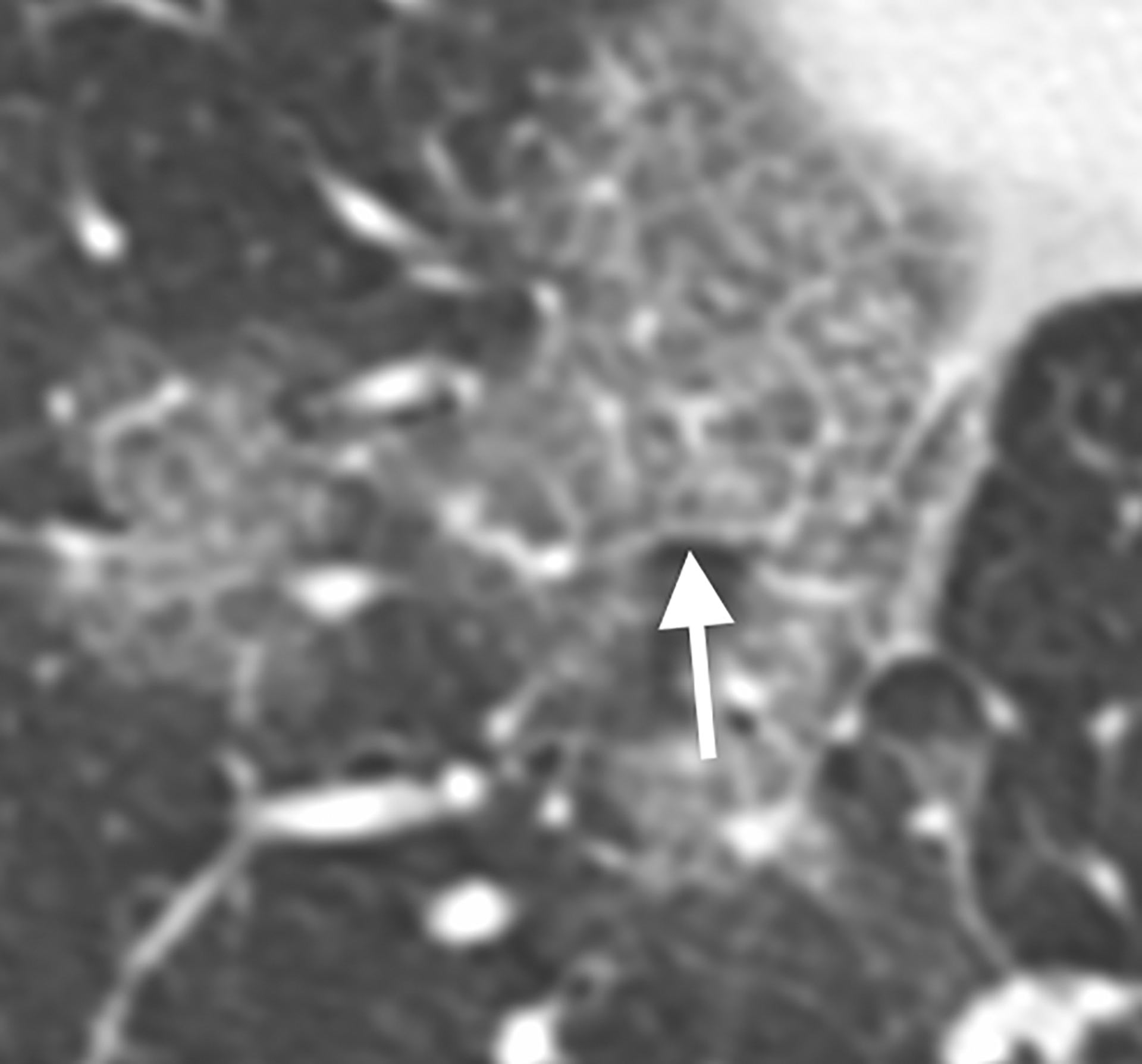
36-year old male with history of recent travel to Wuhan, presenting with fever, fatigue and myalgias. Coronal thin-section non-contrast CT image shows ground-glass opacities with a rounded morphology in both upper lobes (arrows). Image credit: Chung et al, doi: 10.1148/radiol.2020200230.
66-year old female with history of recent travel to Wuhan, presenting with fever and productive cough. Axial thin-section coned-down non-contrast CT image shows a ‘crazy paving’ pattern as manifested by right lower lobe ground-glass opacification and interlobular septal thickening (arrow) with intralobular lines. Image credit: Chung et al, doi: 10.1148/radiol.2020200230.
For each of the 21 patients, the initial CT scan was evaluated for the following characteristics:
(i) presence of ground-glass opacities;
(ii) presence of consolidation;
(iii) number of lobes affected by ground-glass or consolidative opacities;
(iv) degree of lobe involvement in addition to overall lung ‘total severity score;’
(v) presence of nodules;
(vi) presence of a pleural effusion;
(vii) presence of thoracic lymphadenopathy (lymph nodes of abnormal size or morphology);
(viii) presence of underlying lung disease such as emphysema or fibrosis; any other thoracic abnormalities were also noted.
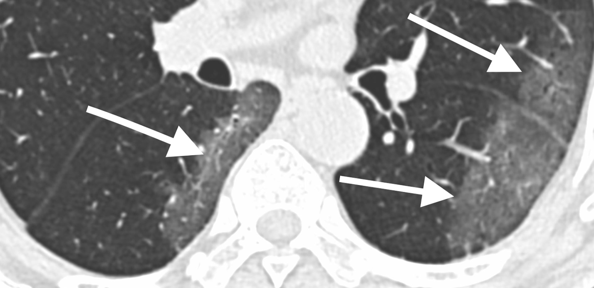
69-year old male with history of recent travel to Wuhan, presenting with fever. Axial thin-section non-contrast CT scan shows ground-glass opacities in the lower lobes with a pronounced peripheral distribution (arrows). Image credit: Chung et al, doi: 10.1148/radiol.2020200230.
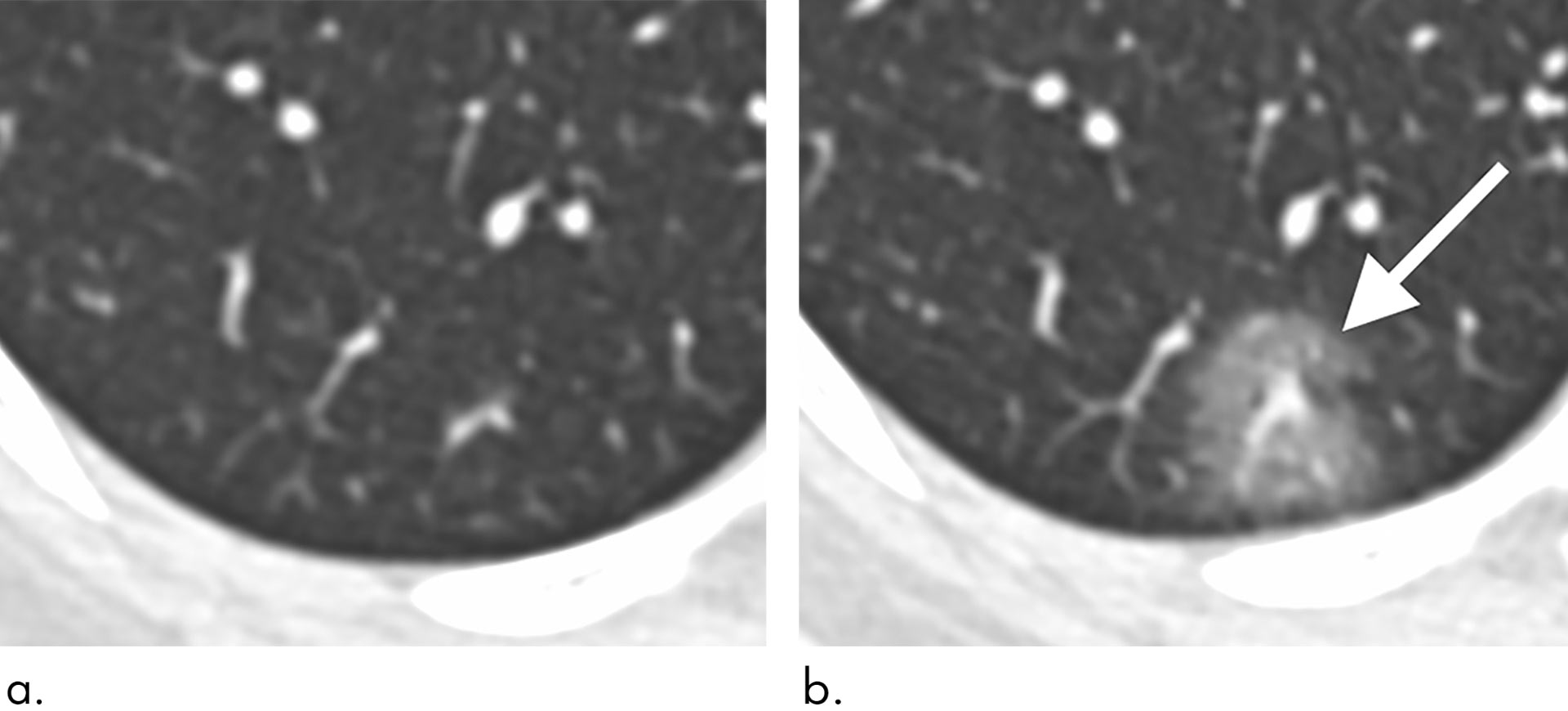
43-year old female with a history of travel to Wuhan presenting with fever: (a) axial thin-section non-contrast CT image from 1/18/2020 shows normal lung; (b) follow-up CT image from 1/21/2020 shows a new solitary, rounded, peripheral ground-glass lesion in the right lower lobe (arrow). Image credit: Chung et al, doi: 10.1148/radiol.2020200230.
The analysis showed that 2019-nCoV typically manifests on CT with bilateral ground-glass and consolidative pulmonary opacities.
Nodular opacities, crazy-paving pattern, and a peripheral distribution of disease may be additional features helpful in early diagnosis.
The researchers also noted that lung cavitation, discrete pulmonary nodules, pleural effusions and lymphadenopathy are characteristically absent in cases of 2019-nCoV.
Follow-up imaging in seven of eight patients showed mild or moderate progression of disease as manifested by increasing extent and density of airspace opacities.
“Absence of abnormal CT findings upon initial examination does not rule out the presence of 2019-nCoV,” Dr. Chung said.
“Our patient population is unique from other published series on the 2019-nCoV coronavirus in that three of our patients had normal initial chest CTs.”
“One of these patients progressed three days later and developed a solitary nodular ground-glass lesion in the right lower lobe, indicating this pattern may represent the very first radiologically visible manifestation of disease in some patients infected with 2019-nCoV.”
“A second patient had a normal follow-up chest CT four days after her initial normal imaging exam.”
“This suggests that chest CT lacks complete sensitivity and does not have a perfect negative predictive value. We can’t rely on CT alone to fully exclude presence of the virus.”
“This finding may be related to the fact that infection with 2019-nCoV is characterized by an incubation period of several days, and there may be a phase where viral infection manifests with symptoms prior to visible abnormalities on CT.”
The team’s report was published online this week in the journal Radiology.
_____
Michael Chung et al. CT Imaging Features of 2019 Novel Coronavirus (2019-nCoV). Radiology, published online February 4, 2020; doi: 10.1148/radiol.2020200230
This article is based on text provided by the Radiological Society of North America.