A team of scientists from Monash University and the Baker Heart and Diabetes Institute has developed a new biosensor that can be used inside a body and is able to emit signals that can be detected by common ultrasound scanners.
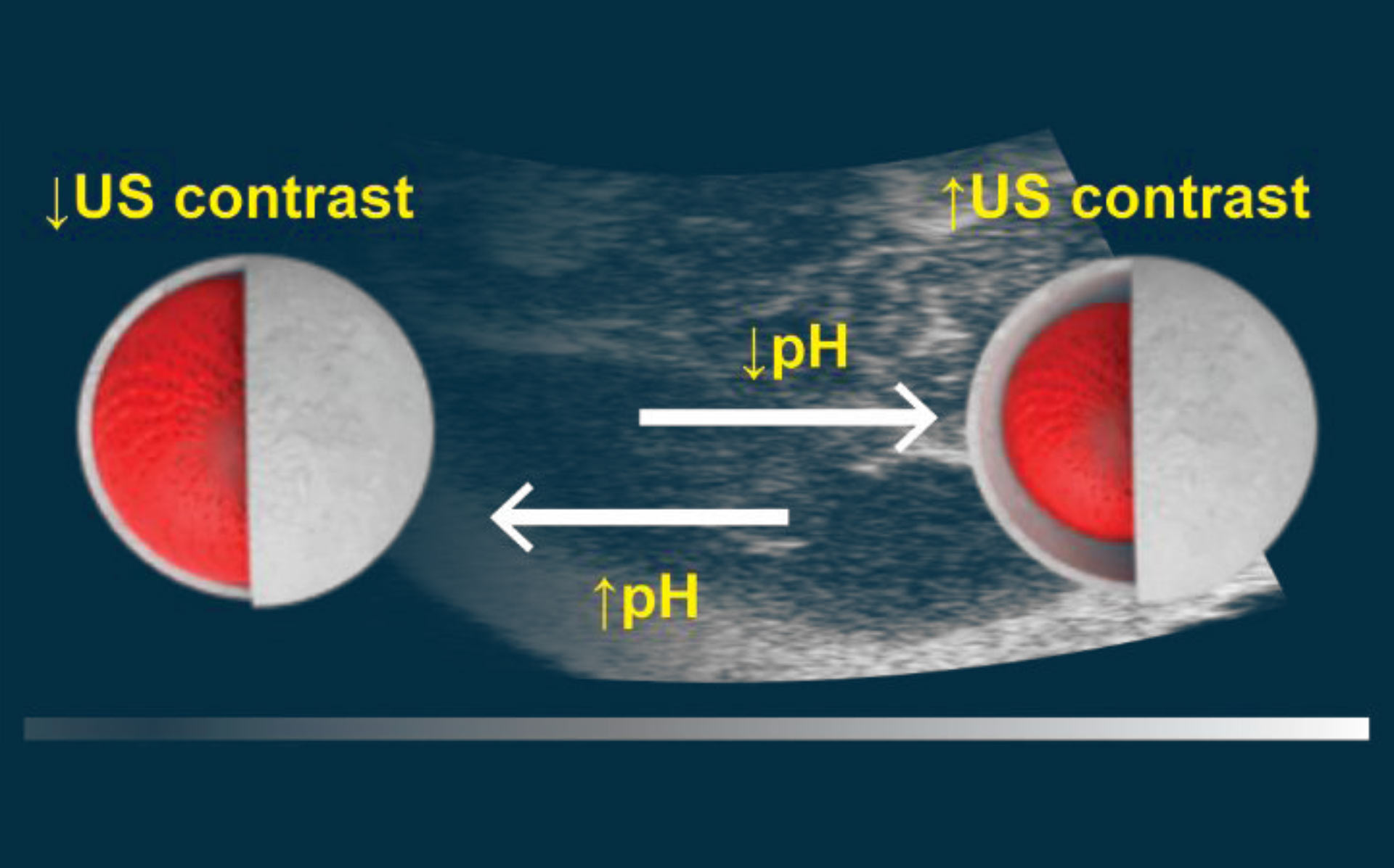
Walker et al developed a solid-state ultrasound contrast agent for monitoring pH fluctuations. Image credit: Walker et al, doi: 10.1021/acssensors.0c00245.
Ultrasound is the most commonly used medical imaging technique as it is portable, non-invasive and allows for tunable tissue penetration.
Tissue discrimination in ultrasound can be enhanced by the use of contrast agents such as commercially available microbubbles, which consist of a gaseous core and a phospholipid shell.
Microbubbles for molecular imaging are a highly attractive field of research, for both diagnostic and theranostic applications. However, these microbubbles have a short imaging time window (5-20 minutes after injection) due to gas diffusion, making them unsuitable for longer-term imaging or feasible for in vivo biosensing studies.
Monash University researchers Dr. Simon Corrie and Dr. Kristian Kempe and their colleagues developed new microbubbles that alter their stiffness in response to pH changes in the body, with these changes picked up by ultrasound.
“Ultrasound imaging uses what is a called a contrast using gas-filled microbubbles,” Dr. Corrie said.
“However, these last only 10-20 minutes making long term tracking within a body impossible.”
The team’s technology can be inserted deep into the tissues and measure biomarkers such as, pH (as a measure of whether a tumor is shrinking following chemotherapy) and in the near future more complex markers such as oxygen (as an indicator of stroke injury) or disease-related proteins.
The advantage of the technology is that, eventually, it will be able to be read by something as simple as a mobile phone which can currently record ultrasound, making it able to monitor patients in remote areas, without the need for big hospital labs.
The technology has been tested in an animal model to detect changes in pH levels. It will now be tested in animal models of disease to determine whether it can accurately monitor rapidly changing pH levels, initially focusing on cancer and stroke.
“The goal is to give clinicians the power of being able to have a patient sit in a chair and, as they are infusing the drugs, using commonly available ultrasound to monitor drug levels or organ response in real-time, adjusting dosages as a function of the patient’s needs,” Dr. Corrie said.
The team’s paper was published in the journal ACS Sensors.
_____
Julia Ann-Therease Walker et al. Dynamic solid-state ultrasound contrast agent for monitoring pH fluctuations in vivo. ACS Sens, published online March 23, 2020; doi: 10.1021/acssensors.0c00245