A team of researchers at the University of Virginia has devised a new imaging approach that combines powerful aspects of both magnetic resonance imaging and gamma-ray imaging.
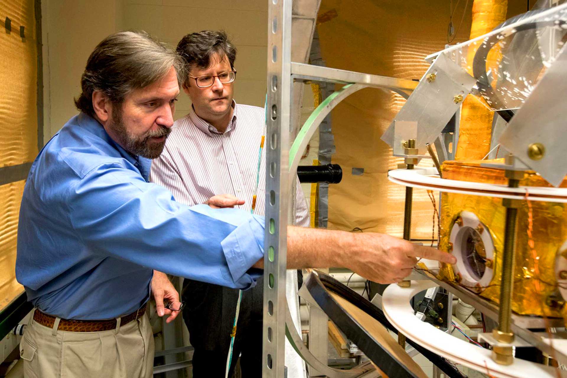
Prof. Gordon Cates and Dr. Wilson Miller with the unique imaging apparatus the team built in their lab. Image credit: Dan Addison / University of Virginia.
The new imaging technique, called polarized nuclear imaging (PNI), has potential for new types of high-resolution medical diagnostics as well as industrial and physics research applications. A paper detailing this technique was published in the Sept. 29, 2016 issue of the journal Nature.
“PNI is a really cool platform technique that fundamentally combined magnetic resonance imaging (MRI) and gamma detection,” said first author Dr. Yuan Zheng, a former graduate student at the University of Virginia.
MRI, which is widely used for detecting cancer and other abnormalities in the body, is effective because it uses a variety of contrast mechanisms to sort out specific characteristics in an image.
And gamma-ray detectors can resolve miniscule amounts of radioactive tracer material, key to homing in on points of particular interest.
“MRI is very powerful in manipulating nuclear spins and generating high resolution images with different contrasts, but it needs lots of nuclear spins to produce a radio-frequency electromagnetic signal that is strong enough to be detected by the receive coils,” explained Dr. Zheng, now with the Department of Radiology at Stanford University.
“While keeping the advantages of MRI, PNI replaces the traditional receive coils by gamma detectors, and measures gamma rays emitted from polarized nuclear tracers with greatly improved sensitivity.”
“With PNI, a tiny amount of radioactive tracers can now be imaged in the ‘MRI’ way, producing much more diagnostic information than current nuclear imaging techniques.”
“This method makes possible a truly new, absolutely different class of medical diagnostics,” added co-author Dr. Wilson Miller, assistant professor of radiology and medical imaging at the University of Virginia.
“We’re combining the advantages of using highly detectable nuclear tracers with the spectral sensitivity and diagnostic power of MRI techniques.”
Dr. Zheng said: “MRI is almost always related to huge equipment. This is because the signal-to-noise ratio of MRI signal depends on the holding magnetic field strength.”
“Expensive, bulky superconducting magnets are usually used to provide this field. Besides, radio-frequency electromagnetic waves from all kinds of daily sources (such as radio stations) may interfere with MRI scans, and could create artifacts in diagnostic images. Therefore MRI scan is usually done in a well-shielded scanner room to get rid of unwanted RF signals.”
“With PNI, things are quite different. The signal-to-noise ratio is independent of the holding field strength, thus superconducting magnets can now be replaced by cheap and lightweight solenoids, just like what we did in our lab.”
“Moreover, PNI depends on the detection of gamma rays instead of radio-frequency signals, which makes RF shielding less critical. Therefore it’s possible to make PNI scanner a portable equipment, and exams don’t have to be done in specially shielded rooms.”
PNI uses magnetic resonance to obtain the spatial information, and then collects image information by detecting gamma rays produced by the tracer material – an isotope of xenon Xe-131m.
“Unlike MRI, which detects faint radio waves, we detect gamma rays that are emitted from the xenon isotope,” explained senior author Dr. Gordon Cates, professor of physics at the University of Virginia.
“Since it is possible to detect a gamma ray from even a single atom, we gain an enormous increase in imaging sensitivity, and dramatically reduce the amount of material needed for performing magnetic-resonance techniques.”
“We chose the metastable isotope Xe-131m mainly because it’s the decay byproduct of commercially available I-131 (a radioactive isotope of iodine for medical use) and is thus readily available to us,” Dr. Zheng added.
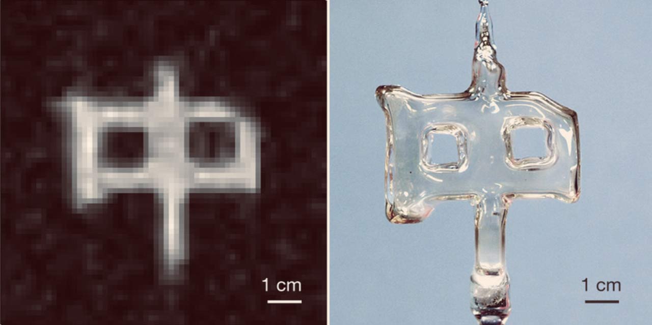
Example of polarized nuclear imaging. Left: image of roughly 1 millicurie of Xe-131m obtained by combining magnetic-resonance techniques with the detection of gamma rays. The image is a 2D projection comprising 32 x 32 pixels, each 3 x 3 mm, and interpolated to 64 x 64 pixels for display. Right: a photograph of the sealed glass cell, shaped like the Chinese character for ‘middle’, in which the sample of Xe-131m was contained. Image credit: Yuan Zheng et al.
The team demonstrated the feasibility of PNI by producing images and spectra from a glass cell containing only about 4 x 10¹³ atoms of Xe-131m.
“We have demonstrated the feasibility of the new technique by producing a proof-of-principle image in a manner never before accomplished,” Prof. Cates noted.
The researchers believe that the technique, once refined, could provide a new, relatively inexpensive way to visualize the gas space of the lungs by having patients inhale a gas containing the isotopes and using PNI to produce an image. The method likewise might work to image targeted areas of the body by injecting isotopes into the bloodstream.
Because PNI would use such small quantities of tracer material, when it comes to medical use, the radioactivity would pose little to no danger to people.
“This technique is in very early stage and it’s difficult to predict exactly what the future medical diagnostic will be like, but we expect the radioactive dose for a PNI exam to be comparable to other nuclear imaging techniques like positron emission tomography (PET),” Dr. Zheng said.
“Considerable work still needs to be done to demonstrate the utility of the new technique in living subjects, but the unique approach represents an exciting new technology,” the scientists said.
“To develop it for practical use, we would need to increase the size of the detectors or the amounts of tracer material, and we are seeking alternative radioactive isotopes that would retain their polarization once inside a living subject.”
“Xe-131m is not the only isotope suitable for PNI,” Dr. Zheng explained.
“Actually, lots of radioactive isotopes can in principle be used for PNI, and some of them may have more favorable properties than Xe-131m, like Kr-79m and Xe-127m, which have shorter half-lives and larger branching ratios for producing the gamma rays of interest.”
_____
Yuan Zheng et al. 2016. A method for imaging and spectroscopy using γ-rays and magnetic resonance. Nature 537, 652-655; doi: 10.1038/nature19775