A group of researchers headed by Brett Hokr of Texas A&M University has demonstrated that a newly emerging technique known as random Raman lasing emission can produce a bright, speckle-free, strobe light source with potential application in high-speed microscopy.

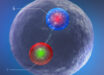
Scientists demonstrated how a narrow-band strobe light source for speckle-free imaging has the potential to reveal microscopic forms of life. Image credit: Research Group Materials for Microelectronic Applications / TU Bergakademie Freiberg.
Random Raman lasing causes a diffuse material such as a powder to emit laser light.
Different from traditional lasers that work by bouncing photons back and forth in a laser cavity, random Raman lasing happens when the light bounces among the powder particles long enough for amplification to occur.
Random Raman laser emission is a pulsed emission with a temporal duration on the scale of single nanoseconds and in a narrow spectrum of about 0.1 nanometer, which can emit a million times more photons per unit time per unit wavelength than any other conventional light source, and should have sufficient intensity to allow scientists to acquire a full two-dimensional fluorescent image in a single pulse of the laser.
“The random Raman laser is unlike any existing laser light source. We found that random Raman lasing emission has a low level of spatial coherence. The emission can be used to produce a wide-field speckle-free quality image with a strobe time on the order of a nanosecond,” Hokr explained.
“This new, bright, fast, narrowband, low-coherence light source opens the door to many exciting new applications in bio-imaging such as high-speed, wide-field microscopy.”
Hokr and his colleagues from Baylor University, TASC Inc., Nanohmics, Inc. and U.S. Air Force Research Laboratory, conducted the first spatial coherence measurement of the random Raman laser in two ways – initially using a classic set-up known as Young’s double slit experiment.
Barium sulfate power was pumped with 530 microjoule, 50 picosecond laser pulses to generate random lasing that later passed through a double slit, and the team captured images of the interference patterns.
The researchers observed that those interference patterns were barely discernible, indicating a very low degree of spatial coherence.
To further quantify the overall spatial coherence, the scientists measured something known as the speckle contrast ratio, which gauges the statistical properties of the emission.
These measurements were consistent in confirming the presence of a low level of coherence.
To further demonstrate that this low coherence truly leads to a speckle-free image, they produced a full-frame, speckle-free microscopic image showing the formation of a cavitation bubble from melanosomes from a several-nanosecond laser pulse at 1,064 nanometer radiation.
The results will be presented May 12 at the CLEO 2015: Conference on Lasers and Electro-Optics in San Jose, California.
_____