An international team of scientists has found alterations of task-based functional brain connectivity in a group of astronauts after a long-duration spaceflight. The findings appear in the journal Frontiers in Physiology.

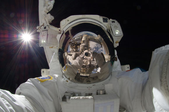
JAXA astronaut Aki Hoshide, ISS Expedition 32 flight engineer, taking a space selfie during extravehicular activity on September 5, 2012, with the Sun behind him. Image credit: NASA.
In the study, Dr. Ekaterina Pechenkova of the HSE Laboratory for Cognitive Research and colleagues compared brain activation and connectivity in two groups: a group of 11 astronauts before and after long-term spaceflight, and healthy controls scanned twice with a comparable interval.
“We used functional magnetic resonance imaging (fMRI) to measure functional brain connectivity in a group of eleven astronauts,” the researchers said.
“We were looking for changes in connectivity between brain areas underlying sensorimotor functions such as movement and perception of body position. These brain areas were activated using gait-imitating plantar stimulation.”
The observed alterations included a disconnection of the vestibular nuclei, the superior part of the right supramarginal gyrus, and the cerebellum from a set of motor, somatosensory, visual, and vestibular areas.
“To compensate for the lack of information from the organs of balance, which cannot provide reliable information in microgravity, the brain develops an auxiliary system of somatosensory control, with increased reliance on visual and tactile feedback instead of vestibular input,” the scientists explained.
“Under Earth’s gravity, vestibular nuclei are responsible for processing signals coming from the vestibular system,” they added.
“But in space, the brain may downweight the activity of these structures to avoid conflicting information about the environment.”
The team also found increased connectivity between the left and right insulae as well as between the part of the right posterior supramarginal gyrus within the temporoparietal junction region and the rest of the brain.
“fMRI showed increased connections between the insular cortex in the left and right hemispheres, as well as between the insular cortex and other areas of the brain,” the study authors said.
“Insular lobes, among other things, are responsible for the integration of signals coming from different sensor systems. Similar functions are performed by the area of parietal cortex in the right supramarginal gyrus, which also demonstrated increased connectivity with other areas of the brain after the flight.”
“It’s an interesting fact that connectivity increase between the right supramarginal gyrus and the left insular cortex was greater among those astronauts who experienced a less comfortable initial adaptation process on the space station (those who experienced vertigo, the illusion of body position, etc.),” Dr. Pechenkova noted.
“We believe this kind of information will eventually help to better understand why it takes different lengths of time for different people to adapt to the conditions of space flight, and will help to develop more effective individual training programs for space travelers,” the researchers concluded.
_____
Ekaterina Pechenkova et al. Alterations of Functional Brain Connectivity After Long-Duration Spaceflight as Revealed by fMRI. Front. Physiol, published online July 4, 2019; doi: 10.3389/fphys.2019.00761


