Astronauts on long-duration flights experience visual impairments due to volume changes in cerebrospinal fluid, the clear fluid that helps cushion the brain and spinal cord while circulating nutrients and removing waste materials, according to a study led by Noam Alperin, professor of radiology and biomedical engineering at the University of Miami Miller School of Medicine.

Astronaut Bruce McCandless II, STS 41-B mission specialist, participates in a historical spacewalk. He is pictured a few meters away from the cabin of the Earth-orbiting Space Shuttle Challenger. This spacewalk represented the first use of a nitrogen-propelled, hand-controlled device called the Manned Maneuvering Unit (MMU), which allows for much greater mobility than that afforded previous space walkers who had to use restrictive tethers. Image credit: NASA.
Over the last decade, NASA scientists began seeing a pattern of visual impairment in astronauts who flew long-duration space missions.
The astronauts had blurry vision, and further testing revealed structural changes of their eyes (papilledema, globe flattening, choroidal folds) and optic nerves (sheath dilation, tortuosity and kinking), as well as signs of elevated intracranial pressure.
The syndrome, known as visual impairment intracranial pressure (VIIP) syndrome, was reported in nearly two thirds of long-duration mission International Space Station (ISS) astronauts.
Researchers previously believed that the primary source of the problem was a shift of vascular fluid toward the upper body that takes place when astronauts spend time in the microgravity of space.
But Prof. Alperin and co-authors recently investigated another possible source for the problem: cerebrospinal fluid (CSF).
The CSF system is designed to accommodate significant changes in hydrostatic pressures, such as when a person rises from a lying to sitting or standing position. However, the microgravity of space presents new challenges.
“On Earth, the CSF system is built to accommodate these pressure changes, but in space the system is confused by the lack of the posture-related pressure changes,” Prof. Alperin explained.
To learn more about the role of CSF in spaceflight-induced visual impairment and eye changes, the team performed high-resolution orbit and brain MRI scans before and shortly after spaceflights for seven long-duration mission ISS astronauts.
The scientists compared results with those from nine short-duration mission space shuttle astronauts.
Using advanced quantitative imaging algorithms, they looked for any correlation between changes in CSF volumes and the structures of the visual system.
The results showed that, compared to short-duration astronauts, long-duration astronauts had significantly increased post-flight flattening of their eyeballs and increased optic nerve protrusion.
Long-duration astronauts also had significantly greater post-flight increases in orbital CSF volume, or the CSF around the optic nerves within the bony cavity of the skull that holds the eye, and ventricular CSF volume — volume in the cavities of the brain where CSF is produced.
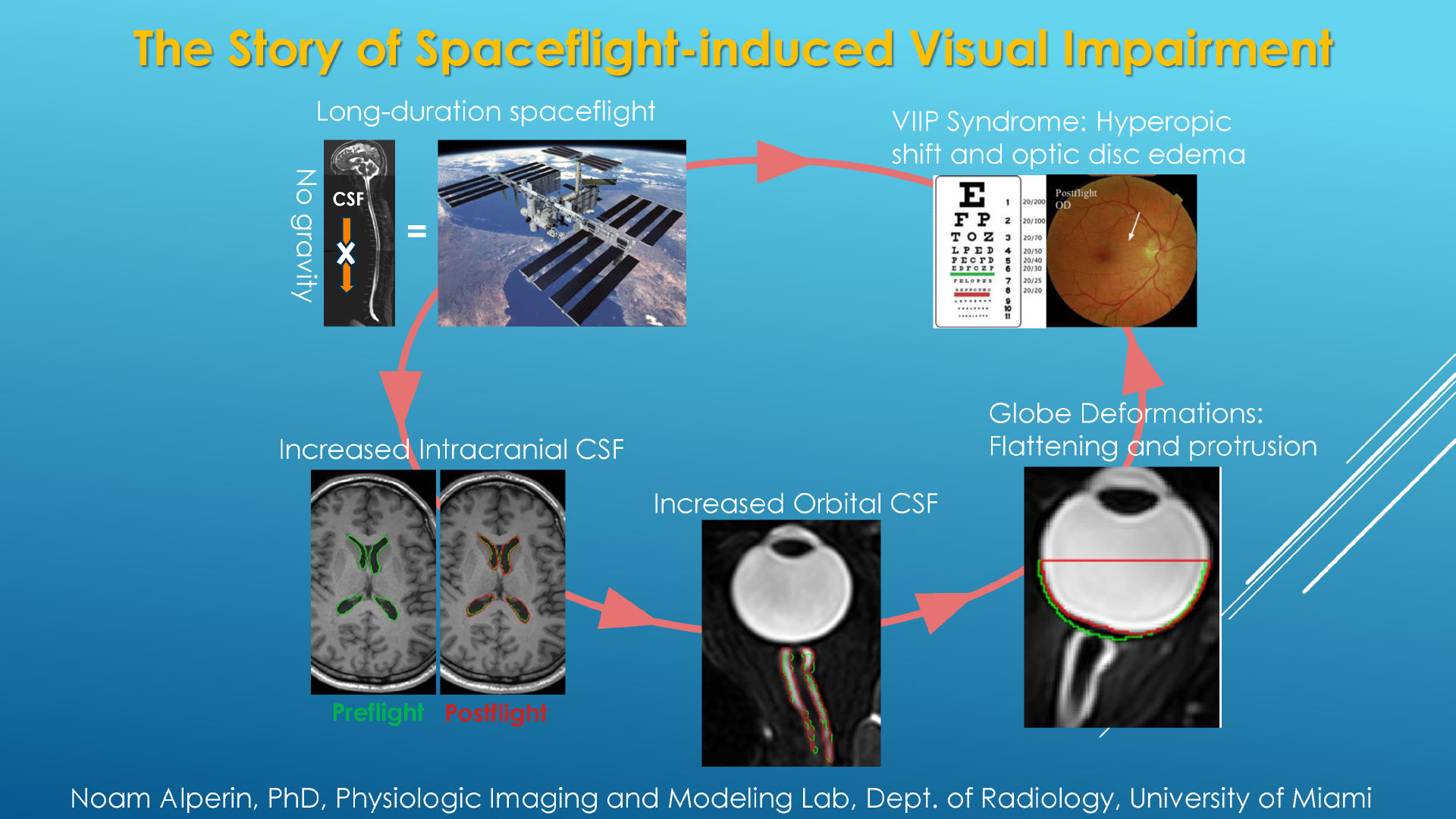
Summary of the spaceflight-induced visual impairment research. Image credit: N. Alperin et al.
“Compared to short-duration astronauts, long-duration astronauts had significantly greater post-flight increases in globe flattening indices (p<0.00001) and optic nerve protrusion indices (p<0.00001),” the authors said.
“Long-duration astronauts also had significantly greater post flight increases in orbital CSF volume (p=0.005) and ventricular CSF volume (p=0.048).”
“There were no significant post flight changes of grey matter volume or white matter volume in either group.”
“The large post spaceflight ocular changes observed in ISS crewmembers were associated with greater increases in intra-orbital and intracranial CSF volume but not with interstitial brain tissue fluid volume.”
This research provides, for the first time, quantitative evidence obtained from short- and long-duration astronauts pointing to the primary and direct role of the CSF in the globe deformations seen in astronauts with visual impairment syndrome.
“Identifying the origin of the space-induced ocular changes is necessary for the development of countermeasures to protect the crew from the ill effects of long-duration exposure to microgravity,” Prof. Alperin said.
“If the ocular structural deformations are not identified early, astronauts could suffer irreversible damage. As the eye globe becomes more flattened, the astronauts become hyperopic, or far-sighted.”
The findings were presented Nov. 28, 2016 at the annual meeting of the Radiological Society of North America in Chicago, IL.
_____
N. Alperin et al. Role of Cerebrospinal Fluid in Spaceflight-Induced Visual Impairment and Ocular Changes. Radiological Society of North America 2016 Scientific Assembly and Annual Meeting, abstract # SSC11-04