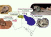
A team of researchers has found a novel retinal structure in the eyes of New World flycatchers. Named the megamitochondria-small oil droplet complex (MMOD complex), this structure may allow these sit-and-wait birds to see their world in a different way from other animals, and help them find and track insect prey more easily.

This light microscopy image of the Acadian flycatcher (Empidonax virescens) retina shows the five traditional oil droplet types and the additional orange, conical structures belonging to the newly described photoreceptor. Image credit: Tyrrell et al, doi: 10.1038/s41598-019-51774-w.
Most birds have four cone photoreceptors for color vision, a fifth cone for non-color-related tasks, and a rod for night vision.
Each cone photoreceptor cell contains a spherical structure called an oil droplet, which filters light before it is converted to electrical signals by the visual pigments, enhancing color discrimination.
Instead of an oil droplet, the MMOD complex — found in two species of New World flycatchers of the genus Empidonax (E. virescens and E. minimus) — contains a high- energy-producing cellular structure called megamitochondria surrounded by hundreds of small, orange-colored droplets.
“We found that Empidonax flycatchers, like all birds, had four single cone photoreceptors that each contained a spherical oil droplet in the inner segment of the photoreceptor,” said senior author Professor Esteban Fernandez-Juricic from the Department of Biological Sciences at Purdue University and colleagues.
“Like other birds, the principal member of the Empidonax double cone also contained a spherical oil droplet. Each type of cone had a different colored oil droplet that could be readily visualized under simple light microscopy.”
“In addition to these five traditional cones and their corresponding oil droplets, we found that the Empidonax flycatcher retina contained what is probably an additional cone photoreceptor with a novel, orange, conical structure in the apical end of the inner segment.”
“Photoreceptors with this organelle lacked the oil droplet that is present in the other cone types.”
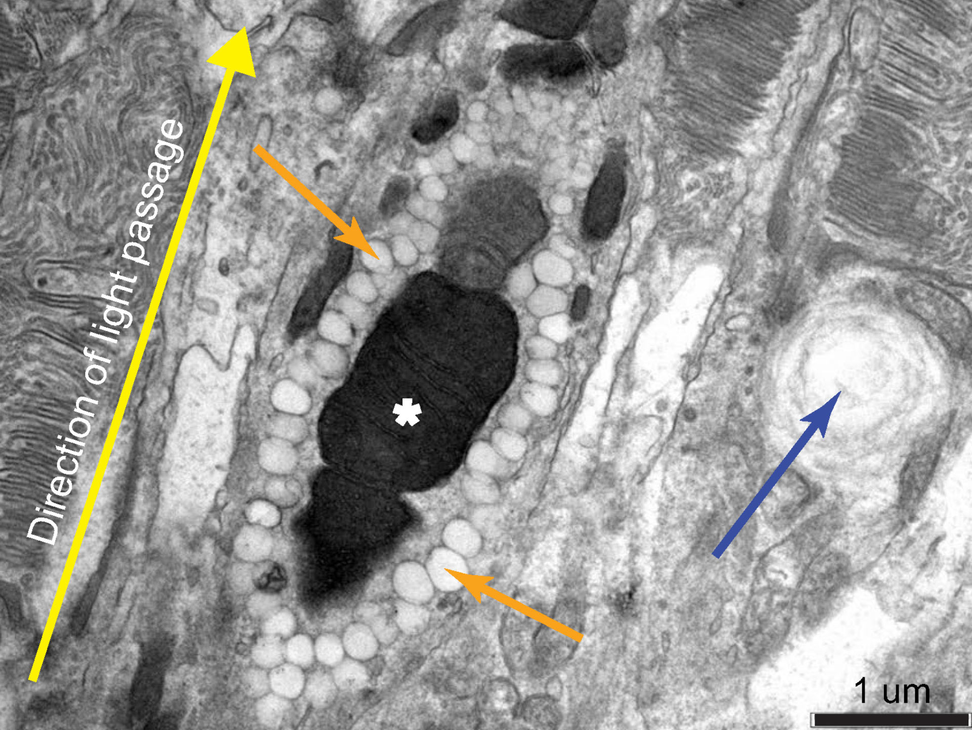
Transmission electron microscopy image of the orange, conical structures reveals that they are electron-dense megamitochondria surrounded by many small oil droplets; the white asterisk denotes the megamitochondria, the orange arrows denote the small oil droplets that provide the orange coloration seen in the image above, and the blue arrow indicates a traditional oil droplet from the neighboring photoreceptor. Image credit: Tyrrell et al, doi: 10.1038/s41598-019-51774-w.
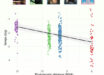
The researchers studied the MMOD complex using light microscopy, transmission electron microscopy, and microspectrophotometry.
They found that this structure works as long-pass filters, letting light with wavelengths longer the 565 nm — or yellow, orange and red — pass through, and absorbing the shorter wavelengths of green, blue and violet.
“The retina of flycatchers, which are sit-and-wait predatory birds, evolved a novel cellular structure in a photoreceptor that may allow them to detect, track and capture fast-moving prey, like insects,” Professor Fernandez-Juricic said.
“This new cone organelle has not been reported before in this form in any other vertebrate retina and may allow these birds to see their world in a different way from other animals,” added Dr. Luke Tyrrell, a researcher in the Department of Biological Science at SUNY Plattsburgh.
The results are published in a paper in the journal Scientific Reports.
_____
L.P. Tyrrell et al. 2019. A novel cellular structure in the retina of insectivorous birds. Sci Rep 9, 15230; doi: 10.1038/s41598-019-51774-w