Chest CT, a routine imaging tool for pneumonia diagnosis, had higher sensitivity for diagnosis of the COVID-19 coronavirus disease as compared with initial reverse-transcription polymerase chain reaction (RT-PCR) from swab samples in the epidemic area of China, according to a paper published in the journal Radiology.
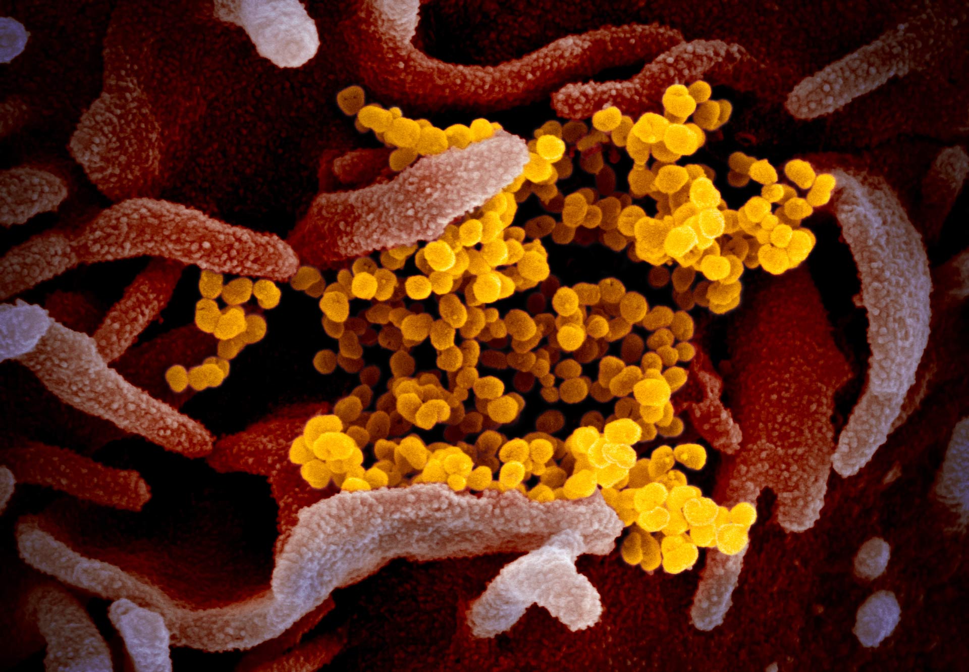
This scanning electron microscope image shows SARS-CoV-2 virus (yellow) isolated from a patient in the U.S., emerging from the surface of cells (pink) cultured in the lab. Image credit: NIAID-RML / CC BY 2.0.
Since December 2019, a number of cases of ‘unknown viral pneumonia’ were reported in Wuhan City, Hubei Province, China.
A novel coronavirus, named SARS-CoV-2, was suspected to be the etiology with Phinolophus bat as the alleged origin.
In just two months, the virus has spread from Wuhan to the whole China, and another 33 countries.
In the absence of specific therapeutic drugs or vaccines for COVID-19, it is essential to detect the disease at an early stage and immediately isolate an infected patient from the healthy population.
According to the latest guidelines published by the Chinese government, the diagnosis of COVID-19 must be confirmed by RT-PCR or gene sequencing for respiratory or blood specimens, as the key indicator for hospitalization.
However, with limitations of sample collection and transportation, as well as kit performance, the total positive rate of RT-PCR for throat swab samples has been reported to be about 30% to 60% at initial presentation.
In the current public health emergency, the low sensitivity of RT-PCR implies that a large number of COVID-19 patients won’t be identified quickly and may not receive appropriate treatment. In addition, given the highly contagious nature of the virus, they carry a risk of infecting a larger population.
“Early diagnosis of COVID-19 is crucial for disease treatment and control. Compared to RT-PCR, chest CT imaging may be a more reliable, practical and rapid method to diagnose and assess COVID-19, especially in the epidemic area,” said senior author Dr. Liming Xia from the Department of Radiology and Tongji Hospital at Huazhong University of Science and Technology Wuhan and colleagues.
Chest CT is fast and relatively easy to perform. Recent research found that the sensitivity of CT for COVID-19 infection was 98% compared to RT-PCR sensitivity of 71%.
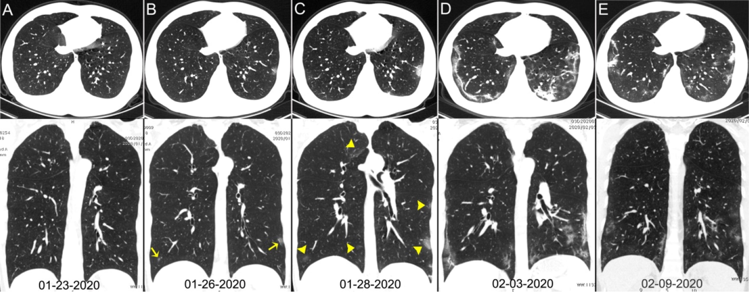
Chest CT images of a 29-year-old man with fever for 6 days; RT-PCR assay for the SARS-CoV-2 using a swab sample was performed on February 5, 2020, with a positive result: (A) normal chest CT with axial and coronal planes was obtained at the onset; (B) chest CT with axial and coronal planes shows minimal ground-glass opacities in the bilateral lower lung lobes (yellow arrows); (C) chest CT with axial and coronal planes shows increased ground-glass opacities (yellow arrowheads); (D) chest CT with axial and coronal planes shows the progression of pneumonia with mixed ground-glass opacities and linear opacities in the subpleural area; (E) chest CT with axial and coronal planes shows the absorption of both ground-glass opacities and organizing pneumonia. Image credit: Ai et al, doi: 10.1148/radiol.2020200642.
For the current study, Dr. Xia and co-authors at Tongji Hospital in Wuhan set out to investigate the diagnostic value and consistency of chest CT imaging in comparison to RT-PCR assay in COVID-19.
The study involved 1,014 patients who underwent both chest CT and RT-PCR tests between January 6 and February 6, 2020.
With RT-PCR as reference standard, the performance of chest CT in diagnosing COVID-19 was assessed.
For patients with multiple RT-PCR assays, the dynamic conversion of RT-PCR test results (negative to positive, and positive to negative, respectively) was also analyzed as compared with serial chest CT scans.
The results showed that 601 patients (59%) had positive RT-PCR results, and 888 (88%) had positive chest CT scans.
The sensitivity of chest CT in suggesting COVID-19 was 97%, based on positive RT-PCR results.
In patients with negative RT-PCR results, 75% (308 of 413 patients) had positive chest CT findings.
Of these, 48% were considered as highly likely cases, with 33% as probable cases.
By analysis of serial RT-PCR assays and CT scans, the interval between the initial negative to positive RT-PCR results was 4 to 8 days.
“About 81% of the patients with negative RT-PCR results but positive chest CT scans were re-classified as highly likely or probable cases with COVID-19, by the comprehensive analysis of clinical symptoms, typical CT manifestations and dynamic CT follow-ups,” the researchers said.
_____
Tao Ai et al. Correlation of Chest CT and RT-PCR Testing in Coronavirus Disease 2019 (COVID-19) in China: A Report of 1014 Cases. Radiology, published online February 26, 2020; doi: 10.1148/radiol.2020200642