A new study, led Boston University researchers, shows that slow oscillating neural activity during non-rapid eye movement (NREM) sleep triggers waves of cerebrospinal fluid that flow in and out of the sleeping brain, washing it of harmful metabolic waste products.

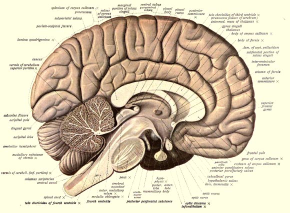
An anatomical illustration from Sobotta’s Human Anatomy, 1908, shows the structure of a human brain. Image credit: Dr Johannes Sobotta.
Sleep is essential for both high-level cognitive processing and also maintenance and restoration of healthy brain function.
The slow-wave neuronal activity of NREM sleep contributes to memory processing and consolidation, whereas cerebrospinal fluid clears metabolic waste products from the brain.
However, it is unknown why these two seemingly unrelated processes co-occur.
In the new study, Dr. Nina Fultz of Boston University and colleagues used accelerated neuroimaging techniques to measure the physiological and neural activity in 11 sleeping human brains.
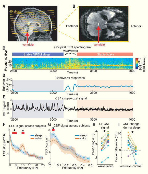
The scientists revealed a pattern between electrophysiological, blood and cerebrospinal fluid dynamics during NREM sleep.
They found that neuronal slow waves are followed by oscillations in blood volume and flow in the brain, which creates space for coupled waves of cerebral spinal fluid to flow in and out of the brain cavity.
“We conclude that human sleep is associated with large coupled low-frequency oscillations in neuronal activity, blood oxygenation, and cerebrospinal fluid flow,” the authors said.
“Although electrophysiological slow waves are known to play important roles in cognition, our results suggest that they may also be linked to the physiologically restorative effects of sleep, as slow neural activity is followed by brain-wide pulsations in blood volume and cerebrospinal fluid flow.”

Large oscillations in cerebrospinal fluid signals appear in the fourth ventricle during sleep. Image credit: Fultz et al, doi: 10.1126/science.aax5440.
“Our results address a key missing link in the neurophysiology of sleep,” they added.
“The macroscopic changes in cerebrospinal fluid flow that we identified are expected to alter waste clearance, as pulsatile fluid dynamics can increase mixing and diffusion. Neurovascular coupling has been proposed to contribute to clearance, but why it would cause higher clearance rates during sleep was not known.”
“Our study suggests slow neural and hemodynamic oscillations as a possible contributor to this process, in concert with other physiological factors.”
“Studies in animals could next test for causal relationships between these neural and physiological rhythms.”
“Our identification of sleep-associated cerebrospinal fluid dynamics also suggests a potential biomarker to be explored in clinical conditions associated with sleep disturbance.”
The results are published in the journal Science.
_____
Nina E. Fultz et al. 2019. Coupled electrophysiological, hemodynamic, and cerebrospinal fluid oscillations in human sleep. Science 366 (6465): 628-631; doi: 10.1126/science.aax5440


