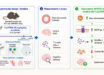
A process using pluripotent stem cells from mice can generate skin tissue complete with hair follicles, according to a team of researchers from the Indiana University School of Medicine. The research is published in the journal Cell Reports.

Dr. Koehler and co-authors describe a defined in vitro 3D culture system that generates skin organoids from mouse pluripotent stem cells. The skin organoids contain self-organized skin layers and skin appendages, including hair follicles, sebaceous glands, and adipocytes. Image credit: Lee et al, doi: 10.1016/j.celrep.2017.12.007.
Although various methods of generating skin tissue in the lab have already been developed, their ability to imitate real skin falls short.
While real skin consists of 20 or more cell types, these models only contain about five or six. Most notably, none of these existing skin tissues is capable of hair growth.
Indiana University School of Medicine’s Dr. Karl Koehler and his colleagues originally began using mouse pluripotent stem cells, which can develop into any type of cells in the body, to create organoids (organs in vitro) that model the inner ear.
But the researchers discovered they were generating skin cells in addition to inner ear tissue, and their research shifted towards coaxing the cells into sprouting hair follicles.
Their recent research demonstrates that a single skin organoid unit developed in culture can give rise to both the epidermis (upper) and dermis (lower) layers of skin, which grow together in a process that allows hair follicles to form the same way as they would in a mouse’s body.
“You can see the organoids with your naked eye. It looks like a little ball of pocket lint that floats around in the culture medium,” Dr. Koehler said.
“The skin develops as a spherical cyst, and then the hair follicles grow outward in all directions, like dandelion seeds.”
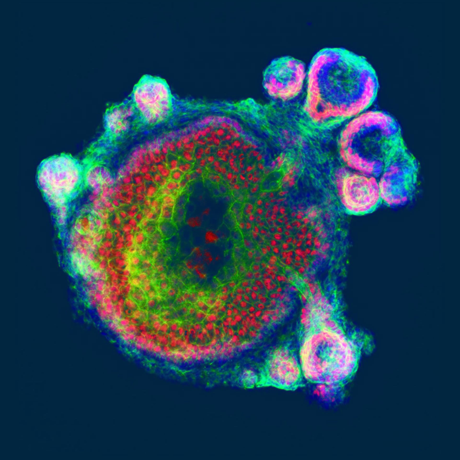
In this artwork, hair follicles grow radially out of spherical skin organoids, which contain concentric epidermal and dermal layers (central structure). Skin organoids self-assemble and spontaneously generate many of the progenitor cells observed during normal development, including cells expressing the protein GATA3 in the hair follicles and epidermis (red). Image credit: Jiyoon Lee / Karl R. Koehler.
While the team was unable to identify exactly which types of hairs developed on the surface of the organoid, they believe the skin grew a variety of hair follicle types similar to those present naturally on the coat of a mouse.
The skin organoid itself consisted of three or four different types of dermal cells and four types of epidermal cells — a diverse combination that more closely mimics mouse skin than previously developed skin tissues.
By observing the development of this more lifelike skin organoid, the authors learned that the two layers of skin cells must grow together in a specific way in order for hair follicles to develop.
As the epidermis grew in the culture medium, it began to take the rounded shape of a cyst. The dermal cells then wrapped themselves around these cysts. When this process was disrupted, hair follicles never appeared.
“It could be potentially a superior model for testing drugs, or looking at things like the development of skin cancers, within an environment that’s more representative of the in vivo microenvironment,” Dr. Koehler said.
“And it would allow us to limit the number of animals we use for research.”
_____
Jiyoon Lee et al. 2018. Hair Follicle Development in Mouse Pluripotent Stem Cell-Derived Skin Organoids. Cell Reports 22 (1): 242-254; doi: 10.1016/j.celrep.2017.12.007