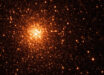
Astronomers using NASA’s Chandra X-ray Observatory have captured the most detailed view yet of the supernova SN 1006.

X-ray image of the SN 1006 remnant. Low, medium, and higher-energy X-rays are colored red, green, and blue respectively (NASA / CXC / Middlebury College / F. Winkler)
The supernova SN 1006 was probably seen first on April 30, 1006, according to records from China and Japan. It was of apparently yellow color, far brighter than Venus and visible during the daytime for weeks.
In the 1960s, astronomers were able to launch instruments and detectors above Earth’s atmosphere to observe the Universe in wavelengths that are blocked from the ground, including X-rays.
SN 1006, located in the constellation of Lupus about 7,200 light years away, was one of the faintest X-ray sources detected by the first generation of X-ray satellites.
In the new Chandra image, low, medium, and higher-energy X-rays are colored red, green, and blue respectively.
The image provides new insight into the nature of the supernova, which is the remnant of a so-called Type Ia supernova.
By examining the different elements in the debris field – such as silicon, oxygen, and magnesium – scientists may be able to piece together how the star looked before it exploded and the order that the layers of the star were ejected, and constrain theoretical models for the explosion.
Scientists are also able to study just how fast specific knots of material are moving away from the original explosion. The fastest knots are moving outward at almost eleven million miles per hour, while those in other areas are moving at a more leisurely seven million miles per hour.
The findings were presented at the 13th Meeting of High Energy Astrophysics Division of the American Astronomical Society in Monterey, CA.
______
Bibliographic information: Winkler PF et al. SN 1006 from Chandra: Exquisite Testament to Progress in Fifty Years of X-ray Astronomy. 13th HEAD Meeting – Monterey, CA. April, 2013; Paper # 400.06
Williams BJ et al. SN 1006 From Chandra: High-resolution Radial Profiles of the Ejecta. 13th HEAD Meeting – Monterey, CA. April, 2013; Paper # 127.09