A new study, published in the journal Development, has confirmed the transient presence of atavistic muscles — present in our ancestors, but normally absent from the adult human — during normal embryonic human development, and reveals the existence of others not previously described in human embryos. Some of these muscles, such as the dorsometacarpales, disappeared from our ancestors more than 250 million years ago, during the transition from synapsid reptiles to mammals. In both the hand and the foot, of the 30 muscles formed at about 7 weeks of gestation one third will become fused or completely absent by about 13 weeks of gestation.

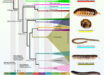
Dorsal view of the left hand of a 10-week old human embryo. The dorsometacarpales are highlighted: these muscles are present in adults of many other limbed animals, while in humans they normally disappear or become fused with other muscles before birth. Image credit: Diogo et al, doi: 10.1242/dev.180349.
Since Charles Darwin proposed his evolutionary theory, scientists have argued that the occurrence of atavistic structures strongly supports the idea that species change over time from a common ancestor through ‘descent with modification.’
For example, ostriches and other flightless birds have vestigial wings, while whales, dolphins and porpoises lack hind limbs but their embryos initiate and then abort hind limb development.
Similarly, temporary small tail-like structures are found in human embryos and the remnant of the lost ancestral tail is retained as our coccyx.
Researchers have also suggested that atavistic muscles and bones can also be seen in human embryos, but it has been difficult to visualize these structures clearly, and the images that appear in modern textbooks are mainly based on decades old analyses.
In the new study, Dr. Rui Diogo and colleagues from Howard University, CNRS and Sorbonne Universites performed the first detailed analysis of the development of human arm and leg muscles using high-quality 3D images of human embryos and fetuses.
The unprecedented resolution of the 3D images revealed the transient presence of several of such atavistic muscles.
The team then compared their observations with few earlier studies that focused on the development of arm and leg muscles in humans, in order to provide information summarizing the timing of appearance, as well as the splitting, fusion and/or loss of each of these muscles.
“It used to be that we had more understanding of the early development of fishes, frogs, chicken and mice than in our own species, but these new techniques allow us to see human development in much greater detail,” said Dr. Diogo, a researcher in the Department of Anatomy at the Howard University College of Medicine.
“What is fascinating is that we observed various muscles that have never been described in human prenatal development, and that some of these atavistic muscles were seen even in 11.5-weeks old fetuses, which is strikingly late for developmental atavisms.”
“Interestingly, some of the atavistic muscles are found on rare occasions in adults, either as anatomical variations without any noticeable effect for the healthy individual, or as the result of congenital malformations,” he said.
“This reinforces the idea that both muscle variations and pathologies can be related to delayed or arrested embryonic development, in this case perhaps a delay or decrease of muscle apoptosis, and helps to explain why these muscles are occasionally found in adult people.”
“It provides a fascinating, powerful example of evolution at play.”
“We hope that our work will not only contribute to the understanding of limb muscle development in humans and in tetrapods in general, but also pave the way for, and stimulate, other researchers to undertake deeper and broader discussions on the links between the upper and lower limbs, between atavisms, variations and anomalies, and between phylogeny and evolution,” the scientists concluded.
_____
Rui Diogo et al. 2019. Development of human limb muscles based on whole-mount immunostaining and the links between ontogeny and evolution. Development 146: dev180349; doi: 10.1242/dev.180349