We would like to think that scientific efforts are free of trends and human whims and proceed in a straight line for the good of humanity; but this is rarely the case, as science like all human affairs is subject to winds of fashion.

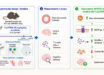
Acute brain slice: thin brain slices allow the selective study of a neural circuit in isolation not possible in intact organism; here one can also selectively manipulate the conditions and observe their specific effect. Image credit: Bhuminder Singh.
Neuroscience is one of the liveliest fields and one that captures the imagination of us all as it aspires to explain who we are, by peering into our minds. Given the excitement, one would think that neuroscience would be witnessing the most judicious use of approaches that would answer questions best for our understanding of brain functioning and neuropsychiatric disorders. This is hardly the case, as can be seen by both the media coverage and journal space occupied by articles that claim to explain our mate, economic and food choices but less so by articles that make tangible progress in detailed understanding of molecules, cells and detailed electrical currents of normal and detailed brain functioning.
These newer, expensive techniques churn out more data and possibly publications, but understanding of the physiology, not so much. Sadly these new techniques also have tendency to pack older more robust techniques into oblivion, rather than accommodating them synergize our learning. There is an acute need to marry the old and the new.
A recent review by Khurana and Li, published in the Reviews in the Neurosciences, is an excellent study and recommendation for an established neurophysiological technique ‘acute brain slice.’ This review begins with evaluating the pros and cons of different approaches in neuroscience. It first drew attention to developments of in vivo multi-electrode recordings, imaging approaches, brain machine interfaces, predictions of behavioral outcomes from neural recordings and problems with many cognitive neuroscience studies. Then it focused on the use of acute brain slices.
Acute brain slices are thin slices of brain (0.1 – 0.5 mm and at time thicker slabs) obtained by decapitating acutely anesthetized animals and after a brief surgery cutting the brain region of interest. Unlike cultured slice in acute slice the neurons are not re-wired, the circuitry is same as in vivo. This is one of the biggest advantages of the technique. Additionally, the electrodes for excitation and recording can be placed with precision. It is easier to access cells and hence both acute and cultured slices are great for understanding the cellular expression patterns of ion channels and all other genes. On the other hand acute slice suffers from damage. What this article provides is insights into minimizing damage and providing ways to recreate in vivo conditions and manipulate them, especially oxygen metabolism that has been ignored by cellular physiologists.
Acute slices have become mainstream after their use by Henry McIlwain’s group in 1960’s for study of ion channels, ion transporters, synaptic functioning and neural computation. Almost all neuropsychiatric diseases that we have some kind of tangible handle on have been studied by extracting living slices of brain and recording from them for few hours to days to figure out the component of molecular and cellular machinery that is defective in case of disease. This success of acute slice in understanding disease related signaling has been due to the ease of access and stable recordings from the exact neurons of defined brain region in question and ability to do pharmacological manipulations of the activity of cells without interference from the global networks.
In recent times the neuropharmacology departments of most pharmaceutical companies have been shut down or drastically reduced due to slow pace of discovery of neuropsychiatric drugs. Instead of picking up the slack, in academia the trend has been to increasingly seek in vivo neurophysiology that is not conducive to mechanistic dissection needed to study molecular amelioration of diseases. Khurana and Li, in this article, capture the problems that would be left unsolved if this migration away from slice continues. Instead of using this article as a reproach against in vivo approaches, these scientists who use both these approaches in their research, provide the best global synthesis on the ideal future of neurophysiology. After pointing to the need of acute slice physiology this article points to concerns that need to be addressed to improve the use of slice physiology. One would think given that acute slices have been used for almost half a decade there would be several articles that discuss pros and cons of acute slice physiology, however, a Pubmed search on systematic evaluation of slice methods reveals none. One would also think that there would have been a more systematic effort to ensure that acute slices are in a state as close to in the intact animal but there has been paucity of that information too.
Apart from providing many existing and novel suggestions on the use of different methods and tools to have minimal damage, recreating brain like cellular interactions, rhythms, this article highlights differences in aerobic metabolism between in vivo and in vitro environment. Khurana and Li make clear suggestions on changes in solutions used in recording, with easy addition of few salts to recreate in vivo like environment. While it remains to be seen if all suggestions of this theoretical study bear fruit, this is the first systematic effort to re-invent slice physiology, a tool that has provided us with most of the neuropsychiatric drugs.
Bhuminder Singh, Ph.D.
(Bhuminder Singh is a research fellow at Vanderbilt University. A cancer researcher by profession, he loves to read about science, its history and evolution as new techniques and ideas integrate with or replace old ones. Twitter: @bhumi_singh).
______
Bibliographic information: Khurana S, Li W. 2013. Baptisms of fire or death knells for acute-slice physiology in the age of ‘omics’ and light? Reviews in the Neurosciences 24 (5): 527-536; doi: 10.1515/revneuro-2013-0028