Radiologists from Wuhan, China found that chest CT had a low rate of missed diagnosis of COVID-19 (3.9%, 2/51) and may be useful as a standard method for the rapid diagnosis of COVID-19 to optimize the management of patients; however, CT remains limited for the identification of specific viruses and distinguishing between viruses.
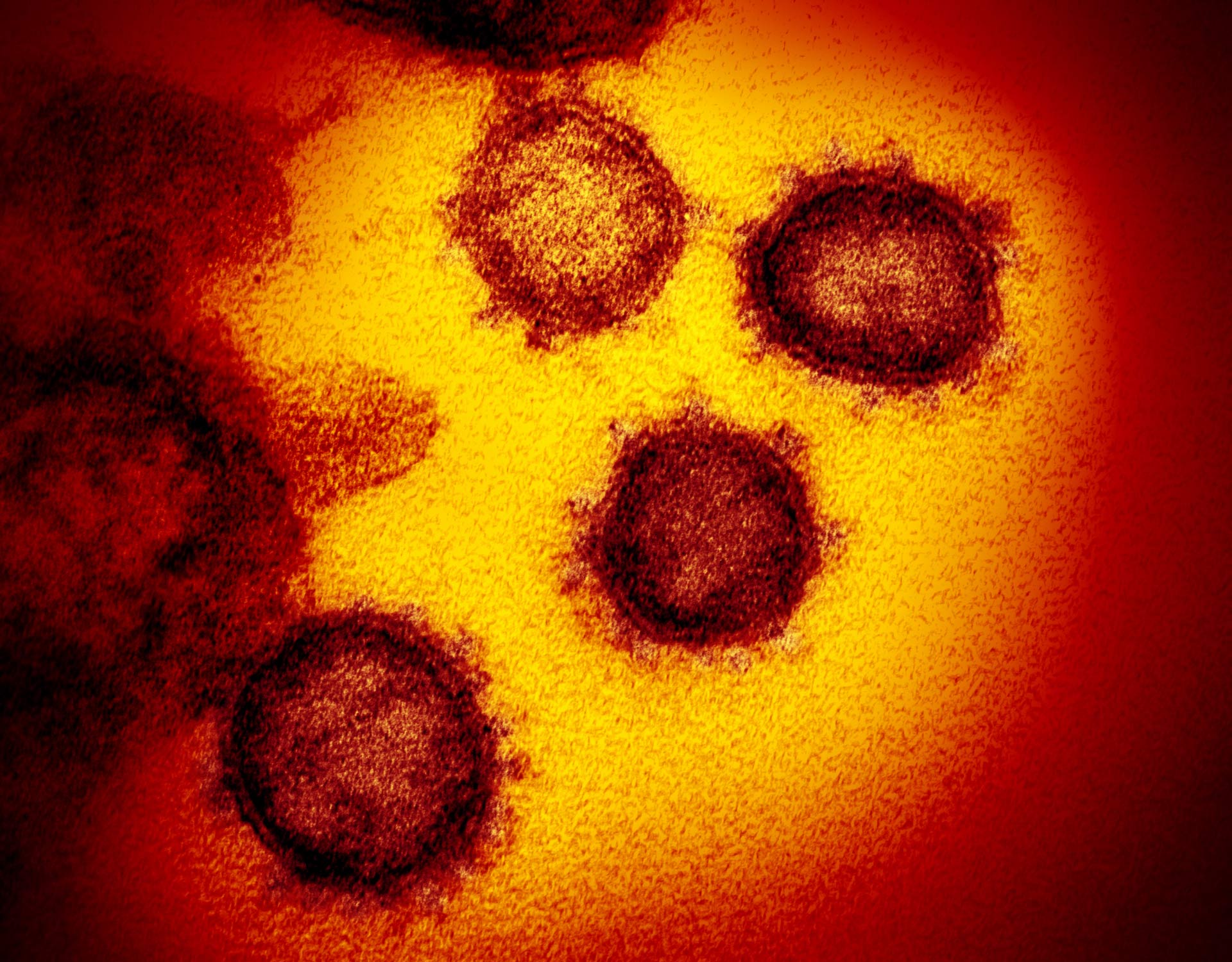
This transmission electron microscope image shows SARS-CoV-2, also known as 2019-nCoV, the virus that causes COVID-19, isolated from a patient in the U.S., emerging from the surface of cells cultured in the lab. Image credit: NIAID-RML.
Yan Li and Liming Xia at Tongji Hospital studied the first 51 patients diagnosed with COVID-19 infection confirmed by nucleic acid testing (23 women and 28 men; age range, 26-83 years) and two patients with adenovirus (one woman and one man; ages, 58 and 66 years).
In their retrospective cohort of 53 patients, as of February 9, 2020, a total of 99 chest CT examinations had been performed.
“Comparing image reports of the initial CT study with laboratory test results to identify patterns suggestive of viral infection, COVID-19 was misdiagnosed as a common infection at the initial CT study in two patients with underlying disease and COVID-19,” the researchers said.
Meanwhile, viral pneumonia was correctly diagnosed at the initial CT study in the remaining 49 patients with COVID-19 and two patients with adenovirus.
“CT of one of the two patients with confirmed adenovirus infection showed ill-defined patchy ground-glass opacities (GGOs), segmental and subpleural consolidations in both lungs, and pleural effusion,” the scientists said.
“CT of the other patient showed subpleural GGOs and consolidation with vascular enlargement, interlobular septal thickening, and air bronchogram sign.”
The CT findings seen in two adenovirus cases were similar to those observed in their COVID-19 cases.
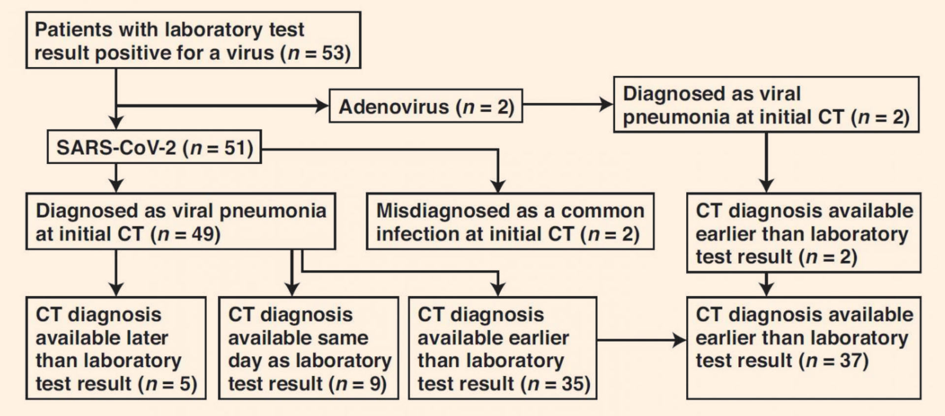
Flowchart shows time difference between positive laboratory test results and positive CT findings for COVID-19 and adenovirus infection in the study group. Image credit: Li & Xia, doi: 10.2214/AJR.20.22954.
The authors also found CT features of COVID-19 that differ from both SARS-CoV and MERS-CoV coronaviruses: a reversed halo sign in two patients (3.9%) and pulmonary nodules with a halo sign in nine patients (17.6%).
“These findings are not mentioned, to our knowledge, in the studies in the literature,” the scientists said.
“It is valuable for radiologists to recognize that the CT findings of COVID-19 overlap with the CT findings of diseases caused by viruses from a different family, such as adenovirus, and have differences as well as similarities with viruses within the same family, such as SARS-CoV and MERS-CoV.”
The team’s paper was published in the American Journal of Roentgenology.
_____
Yan Li & Liming Xia. Coronavirus Disease 2019 (COVID-19): Role of Chest CT in Diagnosis and Management. American Journal of Roentgenology, published online March 4, 2020; doi: 10.2214/AJR.20.22954